. 2002 Feb;109(3):363-71.
doi: 10.1172/JCI13462.

Dipeptidyl peptidase I activates neutrophil-derived serine proteases and regulates the development of acute experimental arthritis

Affiliations

Dipeptidyl peptidase I activates neutrophil-derived serine proteases and regulates the development of acute experimental arthritis

April M Adkison et al. J Clin Invest. 2002 Feb.

Abstract

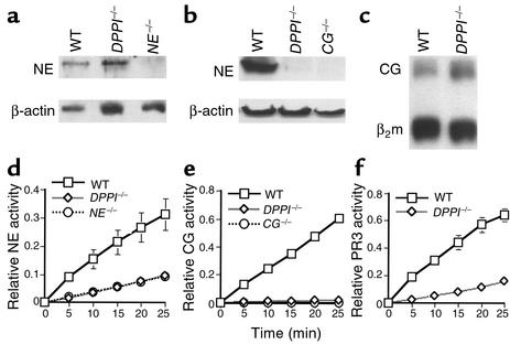
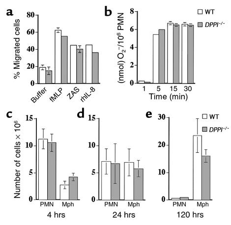
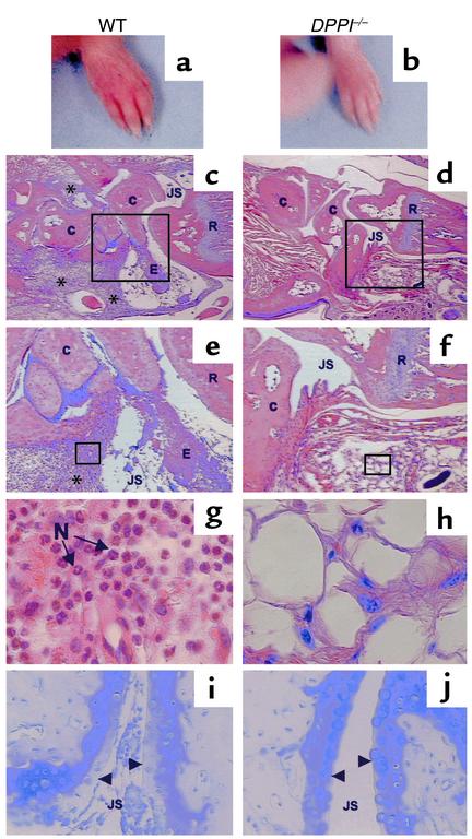
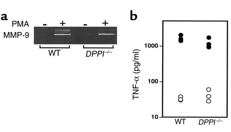
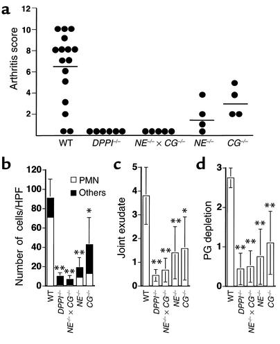
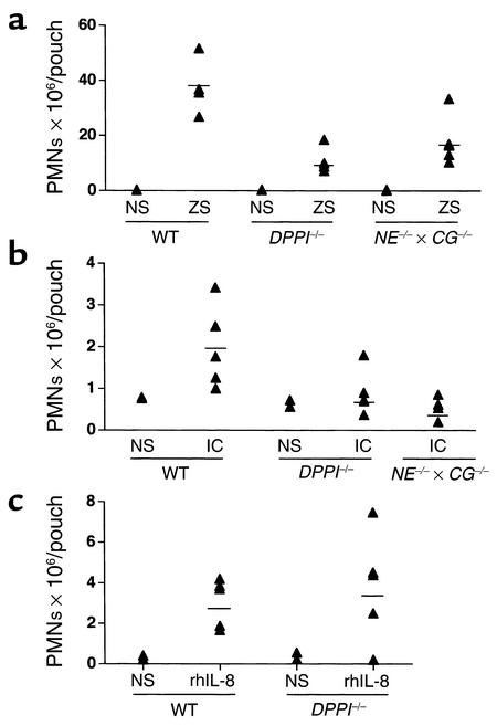
Leukocyte recruitment in inflammation is critical for host defense, but excessive accumulation of inflammatory cells can lead to tissue damage. Neutrophil-derived serine proteases (cathepsin G [CG], neutrophil elastase [NE], and proteinase 3 [PR3]) are expressed specifically in mature neutrophils and are thought to play an important role in inflammation. To investigate the role of these proteases in inflammation, we generated a mouse deficient in dipeptidyl peptidase I (DPPI) and established that DPPI is required for the full activation of CG, NE, and PR3. Although DPPI(-/-) mice have normal in vitro neutrophil chemotaxis and in vivo neutrophil accumulation during sterile peritonitis, they are protected against acute arthritis induced by passive transfer of monoclonal antibodies against type II collagen. Specifically, there is no accumulation of neutrophils in the joints of DPPI(-/-) mice. This protective effect correlates with the inactivation of neutrophil-derived serine proteases, since NE(-/-) x CG(-/-) mice are equally resistant to arthritis induction by anti-collagen antibodies. In addition, protease-deficient mice have decreased response to zymosan- and immune complex-mediated inflammation in the subcutaneous air pouch. This defect is accompanied by a decrease in local production of TNF-alpha and IL-1 beta. These results implicate DPPI and polymorphonuclear neutrophil-derived serine proteases in the regulation of cytokine production at sites of inflammation.


Figures

Figure 1
Figure 1
Analysis of PMN-derived serine proteases in DPPI–/– mice. NE (a) and CG (b) protein expression in DPPI–/– bone marrow lysates. β-actin served as control for protein content. Note the markedly reduced level of immunoreactive CG in DPPI–/– bone marrow lysate compared with WT. (c) S1 nuclease protection assay revealed equivalent levels of CG mRNA in DPPI–/– and WT bone marrow lysate. β2 microglobulin (β2m) served as control for RNA loading. (d) DPPI–/– bone marrow cells and NE–/– bone marrow cells have equivalent residual levels of NE activity. (e) Both DPPI–/– and CG–/– bone marrow cells have no detectable hydrolysis of the CG-specific peptide substrate. (f) Conversion of the PR3 substrate by DPPI–/– bone marrow cell lysate was reduced by 80% compared with WT. Relative enzyme activity was measured at OD405 per 105 bone marrow cells and values represent mean activity ± SEM of at least three animals.
Figure 2
Figure 2
In vitro PMN function and in vivo response to thioglycollate in the absence of DPPI. (a) Bone marrow–derived PMNs from WT and DPPI–/– mice were allowed to migrate across a filter in response to 10–4 M fMLP, 2% zymosan-activated rat serum (ZAS), or 3 μg/ml rhIL-8. The number of cells that migrated was expressed as a percentage of total PMNs. Values represent the mean ± SEM of at least three animals. (b) Superoxide production by PMNs in response to PMA, expressed as nmol of O2 generated per 106 PMNs (n > 3 per genotype). The number of PMNs and macrophages recruited in response to intraperitoneal injection of thioglycollate is normal in DPPI–/– mice at 4 hours (c) 24 hours (d) and 120 hours (e).The slightly lower number of macrophages at 120 hours after thioglycollate injection in the DPPI–/– mice did not reach statistical significance (n = 4–5 mice per genotype). Mph, macrophages.
Figure 3
Figure 3
Clinical and histological assessment of monoclonal antibody–induced arthritis. Forepaw of a WT mouse (a) and a DPPI–/– mouse (b) on day 7 after receiving arthrogenic antibodies against type II collagen. Note the redness and swelling involving the entire paw, including the digits, of the WT mouse (a). Histological analysis of sections taken from the radiocarpal joints of immunized animals, stained with hematoxylin and eosin (ch), and toluidine blue (i and j) (to detect proteoglycan). WT mice (c, e, g, and i) have marked leukocyte infiltration (*) in the subsynovial tissue and joint space, adherence of inflammatory cells to joint surfaces, and proteoglycan loss as evidenced by the reduction in metachromasia at the superficial border of the cartilage matrix (arrowheads). The infiltrating cells are predominantly neutrophils (N) (g). DPPI–/– mice were protected from arthritis, as evidenced by normal joint histology, lack of inflammatory infiltrates, and preservation of proteoglycan content (d, f, h, and j). Magnification: c and d, ×16; e and f, ×40; g and h, ×400. E, exudate; C, carpal bones; R, radius; JS, joint space.
Figure 4
Figure 4
In vitro neutrophil degranulation and TNF-α production in DPPI–/– mice. (a) Purified PMNs (107/ml) were incubated with 50 ng/ml PMA for 15 minutes, and cell-free supernatants were analyzed by gelatin zymography. Note that DPPI–/– and WT PMNs released equivalent levels of MMP-9. (b) TNF-α production 90 minutes after intraperitoneal injection of 100 μg LPS. Open circles represent TNF-α levels prior to LPS injection. Serum TNF-α levels (filled circles) after LPS injection were equivalent in WT mice and DPPI–/– mice.
Figure 5
Figure 5
Summary of clinical arthritis and histological scores of immunized mice. (a) Groups of mice with different genotypes were immunized with arthrogenic antibodies on day 0, followed by LPS on day 3. Each paw was evaluated and scored on a scale of 0–3, as outlined in Methods. Results are expressed as the mean arthritis score on day 7 of all four paws (maximum score of 12 per mouse). Joint sections were evaluated by a blinded observer for cellular infiltrate (b), severity of joint exudate (c), and proteoglycan (PG) depletion(d). Degree of proteoglycan depletion was scored on a scale of 0–3 with 3 being most severe. Results are expressed as mean ± SEM for each of the histological features, quantified in WT, DPPI–/–, NE–/– × CG–/–, NE–/–, and CG–/– mice (n = at least four animals and 16 paws per genotype). *P < 0.01, **P < 0.001.
Figure 6
Figure 6
Neutrophil recruitment in air pouch model. Neutrophil recruitment in response to zymosan (ZS) (a), immune complex (IC) formation (b), and rhIL-8 (c). Mice were sacrificed 12 hours after injection of ZS and IC formation and 4 hours after rhIL-8. Each point represents the number of PMNs in an individual pouch. NS, normal saline.

References

    1. Lehrer RI, Ganz T. Antimicrobial polypeptides of human neutrophils. Blood. 1990;76:2169–2181. - PubMed
    1. Chertov O, Yang D, Howard OM, Oppenheim JJ. Leukocyte granule proteins mobilize innate host defenses and adaptive immune responses. Immunol Rev. 2000;177:68–78. - PubMed
    1. Weiss SJ. Tissue destruction by neutrophils. N Engl J Med. 1989;320:365–376. - PubMed
    1. Lehr H, Arfors K. Mechanisms of tissue damage by leukocytes. Curr Opin Hematol. 1994;1:92–99. - PubMed
    1. Caughey GH. Serine proteinases of mast cell and leukocyte granules. A league of their own. Am J Respir Crit Care Med. 1994;150:S138–S142. - PubMed

Publication types

MeSH terms