. 2002 Feb 4;195(3):343-51.
doi: 10.1084/jem.20011149.

Human dendritic cells activate resting natural killer (NK) cells and are recognized via the NKp30 receptor by activated NK cells

Affiliations

Human dendritic cells activate resting natural killer (NK) cells and are recognized via the NKp30 receptor by activated NK cells

Guido Ferlazzo et al. J Exp Med. .

Abstract

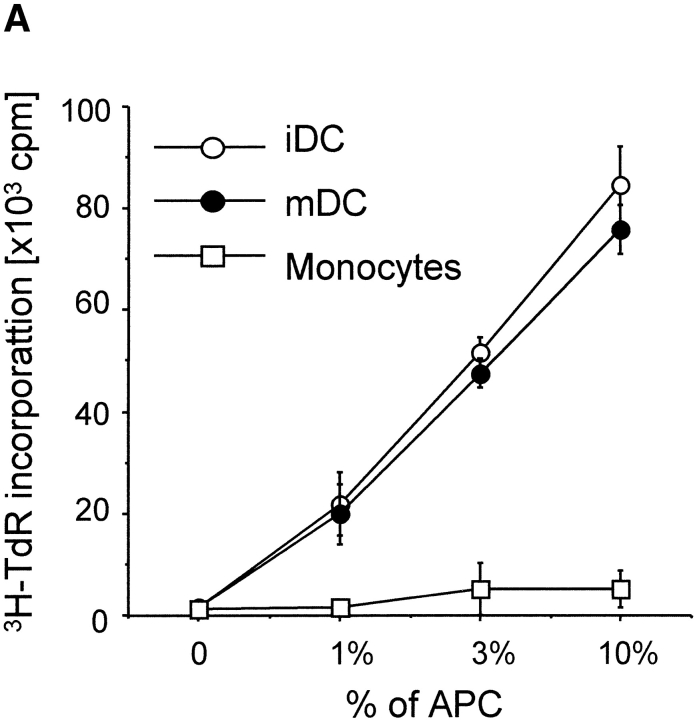
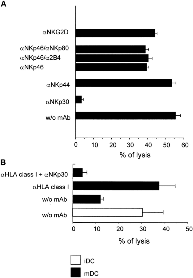
During the innate response to many inflammatory and infectious stimuli, dendritic cells (DCs) undergo a differentiation process termed maturation. Mature DCs activate antigen-specific naive T cells. Here we show that both immature and mature DCs activate resting human natural killer (NK) cells. Within 1 wk the NK cells increase two-- to fourfold in numbers, start secreting interferon (IFN)-gamma, and acquire cytolytic activity against the classical NK target LCL721.221. The DC-activated NK cells then kill immature DCs efficiently, even though the latter express substantial levels of major histocompatibility complex (MHC) class I. Similar results are seen with interleukin (IL)-2--activated NK cell lines and clones, i.e., these NK cells kill and secrete IFN-gamma in response to immature DCs. Mature DCs are protected from activated NK lysis, but lysis takes place if the NK inhibitory signal is blocked by a human histocompatibility leukocyte antigen (HLA)-A,B,C--specific antibody. The NK activating signal mainly involves the NKp30 natural cytotoxicity receptor, and not the NKp46 or NKp44 receptor. However, both immature and mature DCs seem to use a NKp30 independent mechanism to act as potent stimulators for resting NK cells. We suggest that DCs are able to control directly the expansion of NK cells and that the lysis of immature DCs can regulate the afferent limb of innate and adaptive immunity.


Figures

Figure 1.
Figure 1.
DCs stimulate proliferation and expansion of NK cells. (A) Small numbers of mature (mDC) and immature DCs (iDC) stimulate proliferation of negatively selected, peripheral blood NK cells, whereas monocytes had little or no stimulating activity. (B) Immature and mature DCs expand NK cell numbers by two- to fourfold, whereas monocytes sustain the initial number of added NK cells (105). Count of viable cells was performed by trypan blue exclusion. Similar results were obtained in five independent experiments.
Figure 1.
Figure 1.
DCs stimulate proliferation and expansion of NK cells. (A) Small numbers of mature (mDC) and immature DCs (iDC) stimulate proliferation of negatively selected, peripheral blood NK cells, whereas monocytes had little or no stimulating activity. (B) Immature and mature DCs expand NK cell numbers by two- to fourfold, whereas monocytes sustain the initial number of added NK cells (105). Count of viable cells was performed by trypan blue exclusion. Similar results were obtained in five independent experiments.
Figure 2.
Figure 2.
The phenotype of DCs after coculture with autologous freshly isolated NK cells. NK cells and DCs were cultured under the same conditions described in Materials and Methods for proliferation assay. The NK/DC ratio in the coculture was 10:1. Surface expression of two DC maturation markers, CD25 (A) and CD83 (B), is shown for DCs gated either as large (FSC, A) or CD11c+ (B) cells. Left column: phenotype of the immature (iDC) and mature DCs (mDC) added to the NK/DC cocultures. Mature DCs were all positive for CD11c and CD83, and 70–80% were CD25high. Immature DCs had low expression of CD83 and CD25. Middle column: immature and mature DCs after 7 d without NK cells. Right column: immature and mature DCs after 7 d with NK cells. Around 10% of immature DCs expressed the mature DC phenotype upon NK coculture. These data are representative of two experiments.
Figure 3.
Figure 3.
DCs induce cytolytic function and IFN-γ secretion by DC-activated NK cells isolated from blood. (A) NK cells were cocultured with immature (iDC) or mature DCs (mDC) for 7 d (the two panels to the left) and then tested for cytolytic activity against DCs and the standard NK target, LCL721.221. The latter and immature DCs were lysed comparably. In contrast, NK cells cocultured with monocytes did not develop any cytolytic activity. (B) NK cells secreted IFN-γ in response to immature and mature DCs, but not monocytes. Similar results after 7 d coculture were obtained in three experiments. The E/T ratio in these experiments was 30:1.
Figure 4.
Figure 4.
NK-mediated lysis of mature vs. immature DC. IL-2–activated autologous NK cells were tested in standard 51Cr release assay at various E/T ratios. Both mature (mDC) and immature DC (iDC) were analyzed. MHC class I LCL721.221 cells were used as an NK-sensitive control. The data shown were obtained with a polyclonal NK population cultured for 20 d in the presence of IL-2 (mean of triplicates).
Figure 5.
Figure 5.
Role of activating NK receptors and coreceptors in the lysis of DCs. (A) IL-2–activated NK cells were analyzed for their cytolytic activity against autologous immature DCs in the absence or in the presence of the indicated mAb. The E/T ratio was 20:1. The experiment shown is representative of six independent experiments and data are mean of triplicates. (B) Black bars represent lysis of mature DCs; the white bar refers to the lysis of immature DCs. Mature DCs display a higher susceptibility to NK-mediated lysis after coating with anti-HLA class I mAb (A6136, IgM) and NKp30 plays a major role in this lysis. The E/T ratio was 20:1. Data are representative of two independent experiments performed in triplicates.
Figure 6.
Figure 6.
Activated NK cells produce IFN-γ upon interaction with autologous immature DCs. Polyclonal (A) or clonal (B) NK cell populations were cocultured with DCs in the absence or in the presence of the indicated mAbs. The E/T ratio was 20:1. The K562 cell line was used as positive control, IFN-γ in the supernatants was detected by ELISA after 48 h. iDC: immature DCs; NK bulk: polyclonal NK population; clone 38: a representative NK cell clone; iDC + NK: NK cells cocultured with DCs alone or in the presence of the indicated mAbs. Data shown are the mean of triplicates. Similar results were obtained in five independent experiments.
Figure 6.
Figure 6.
Activated NK cells produce IFN-γ upon interaction with autologous immature DCs. Polyclonal (A) or clonal (B) NK cell populations were cocultured with DCs in the absence or in the presence of the indicated mAbs. The E/T ratio was 20:1. The K562 cell line was used as positive control, IFN-γ in the supernatants was detected by ELISA after 48 h. iDC: immature DCs; NK bulk: polyclonal NK population; clone 38: a representative NK cell clone; iDC + NK: NK cells cocultured with DCs alone or in the presence of the indicated mAbs. Data shown are the mean of triplicates. Similar results were obtained in five independent experiments.

References

    1. Banchereau, J., and R.M. Steinman. 1998. Dendritic cells and the control of immunity. Nature. 392:245–252. - PubMed
    1. Thery, C., and S. Amigorena. 2001. The cell biology of antigen presentation in dendritic cells. Curr. Opin. Immunol. 13:45–51. - PubMed
    1. Banchereau, J., F. Briere, C. Caux, J. Davoust, S. Lebecque, Y.J. Liu, B. Pulendran, and K. Palucka. 2000. Immunobiology of dendritic cells. Annu. Rev. Immunol. 18:767–811. - PubMed
    1. Ljunggren, H.G., and K. Karre. 1985. Host resistance directed selectively against H-2-deficient lymphoma variants. Analysis of the mechanism. J. Exp. Med. 162:1745–1759. - PMC - PubMed
    1. Ljunggren, H.G., and K. Karre. 1990. In search of the ‘missing self’: MHC molecules and NK cell recognition. Immunol. Today. 11:237–244. - PubMed

Publication types

MeSH terms