. 2002 Feb;129(3):551-61.
doi: 10.1242/dev.129.3.551.

Tail gut endoderm and gut/genitourinary/tail development: a new tissue-specific role for Hoxa13

Affiliations

Tail gut endoderm and gut/genitourinary/tail development: a new tissue-specific role for Hoxa13

Pascal de Santa Barbara et al. Development. 2002 Feb.

Abstract

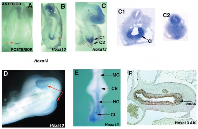
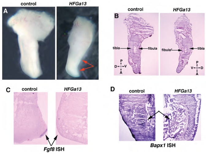
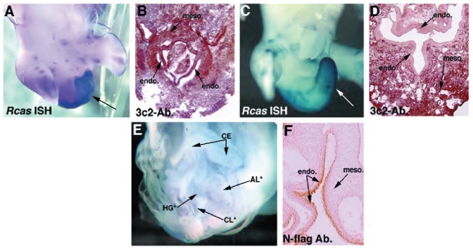
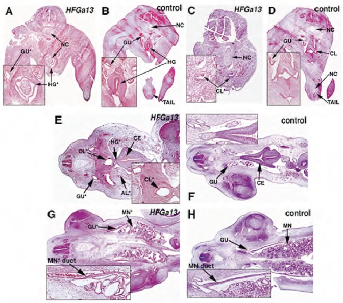
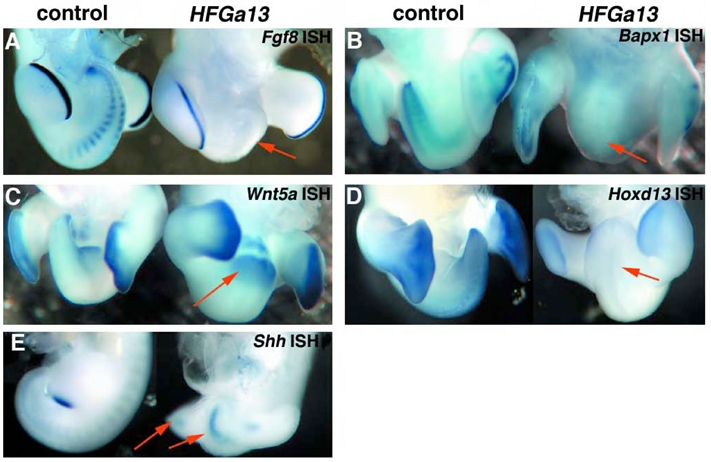
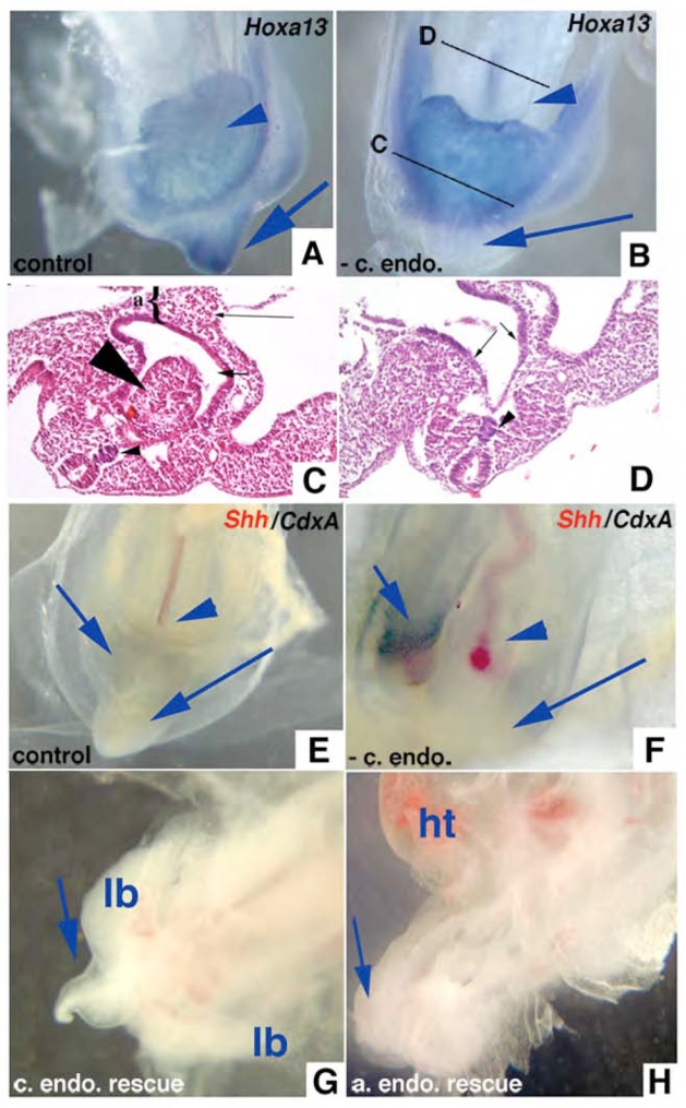
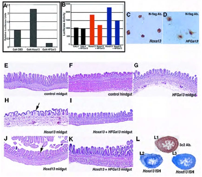
Hoxa13 is expressed early in the caudal mesoderm and endoderm of the developing hindgut. The tissue-specific roles of Hoxa13 function have not been described. Hand-foot-genital syndrome, a rare dominantly inherited human malformation syndrome characterized by distal extremity and genitourinary anomalies, is caused by mutations in the HOXA13 gene. We show evidence that one specific HOXA13 mutation likely acts as a dominant negative in vivo. When chick HFGa13 is overexpressed in the chick caudal endoderm early in development, caudal structural malformations occur. The phenotype is specific to HFGa13 expression in the posterior endoderm, and includes taillessness and severe gut/genitourinary (GGU) malformations. Finally, we show that chick HFGa13 negatively regulates expression of Hoxd13 and antagonizes functions of both endogenous Hoxa13 and Hoxd13 proteins. We suggest a fundamental role for epithelial specific expression of Hoxa13 in the epithelial-mesenchymal interaction necessary for tail growth and posterior GGU patterning.


Figures

Fig. 1
Fig. 1
Spatio-temporal expressions of Hoxa13 (A–E) and Hoxa13 protein (F) during posterior GGU/tail development. (A) St. 10 (arrow at CIP). (B) St. 14 (arrow at CIP). (C) St. 17 (long arrows show tail tip, short arrow at hindgut and arrowhead at tailgut). Planes of cryosection are indicated by short arrows (C1, C2). Hoxa13 endodermal expression in the cloaca (CL). (D) expression in the tail and hindlimb bud at St. 22. (E) dissected E4 posterior gut (arrows at endodermal hindgut (HG) and mesodermal cloacal expression), no expression is detected in the ceca (CE) or midgut (MG). (F) protein expression at E6 section of the hindgut in the endoderm (endo) hindgut (arrow).
Fig. 2
Fig. 2
Human HFG and chick HFGa13 overexpressed embryos have similar limb phenotypes. (A–D) Control (left panel) and HFGa13-infected (right panel) hindlimbs at E6. Planar sections of E6 limbs (B), uninjected control (left) HFGa13 infected hindlimb (right, arrow shows fibula maldevelopment with undifferentiated mesenchymal cells). (C) In situ hybridization of Fgf8 in sectioned hindlimb. No Fgf8 expression is detected in the HFGa13-infected hindlimb compare to the uninfected hindlimb (arrows). (D) In situ hybridization of Bapx1 in sectioned hindlimb. Altered Bapx1 expression in the HFGa13-infected hindlimb (arrows).
Fig. 3
Fig. 3
Endodermally expressed HFGa13 causes abnormal hindgut and tail development. (A) Whole-mount in situ hybridization showing RCAS expression in an E6 mutant HFGa13 infected embryo. (B) 3C2 immunohistochemistry analysis of sectioned HFGa13-infected mutant embryo shows posterior endoderm (endo.) and mesoderm (meso.) infection. (C) RCAS in situ showing absence of the mutant phenotype when HFGa13 infection is present only in the mesoderm, demonstrated in (D) with anti-gag immunohistochemistry showing no infection in the caudal endoderm (E6). (E) E6 survivors after electroporation of HFGa13 constructs in the posterior endodermal layer. The phenotype involves maldevelopment of the cloaca (CL*), hindgut (HG*), and tail. Allantoic internalization is present (AL*), ceca are unaffected (CE). (F) Anti-Nflag demonstrating expression of the tagged-HFGa13 in the endoderm of the hindgut, mesoderm is not stained. Note the electroporated endodermal cells appear undamaged and intact. Misexpression of HFGa13 and Hoxa13 constructs by electroporation show similar expression levels in the gut endoderm layer (data not shown).
Fig. 4
Fig. 4
Histology analysis of mutant HFGa13-infected embryos. H&E stained transverse (A–D, E7) and longitudinal (E–H, E6) sections of mutant HFGa13-infected (A,C,E,G) and control embryos (B,D,F,H)- Ourentery is present in (A). HFGa13-infected embryos show cloacal stenosis (C), atresia of the hindgut anterior to the cloaca (E), allantoic internalization (E), and defects in the cloaca-associated viscera including more distal Müllerian duct (MN duct) atresia and cystic mesonephric (MN*) maldevelopment (G). Note the correct development of the more anterior gut structures. Ceca (CE), notochord (NC), other abbreviations as per Fig. 3.
Fig. 5
Fig. 5
HFGa13 affects expression of Fgf8, Hoxd13 and Bapx1. (A–G) Whole-mount in situ hybridizations of control (left panel) and HFGa13-infected (right panel) embryos (arrows at malformed tail). (A) Fgf8 expression at E5, note absence of expression in the HFGa13 tail but normal expression in the hindlimb AER. (B) Bapx1 (expression) at E6. Bapx1 shows a diminished expression in the tail of the HFGa13-infected embryo, but normal expression in the non-infected hindlimbs. (C) Wnt5a gene expression shows expression in the HFGa13-infected embryo, E7. (D) Hoxd13 expression at E6 shows strong down-regulation in the tail HFGa13-infected embryo and normal expression in hindlimbs. (E) Shh expression at E4 shows is normal. Arrows at hindlimb and notochord.
Fig. 6
Fig. 6
Whole embryo explants developed in culture demonstrate the physical necessity of caudal endoderm for normal tail development. (A) Whole embryo explanted at St. 10 and grown on albumen/agarose for 48 hours showing normal tail development and expression of Hoxa13 in tail (blue arrow). Arrowhead shows normal placement of allantois ventral to hindgut which expresses Hoxa13. (B) St. 10 embryo grown with caudal endoderm removed demonstrating blunted tail without Hoxa13 expression (long arrow). Short arrow marks hindgut dorsal to Hoxa13 expressing allantois. (C) H&E section through a caudal endoderm-less embryo that developed ourentery (large arrowhead) within hindgut lumen (short arrow). {shows allantois. Small arrowhead shows normal notochord. (D) Section anterior to (C) showing endoderm (arrows) and notochord (arrowhead). (E) Embryo cultured ~24 hours with caudal endoderm develops normal CIP (long arrow), expresses Shh in notochord (red stain, arrowhead, most of notochord deep to plane of photograph), and coexpresses Shh and CdxA in endoderm of CIP (short arrow, purple/black stain). (F) Embryo cultured ~24 hours without caudal endoderm fails to develop CIP or express Shh or CdxA in midline caudal endoderm (long arrow), both are coexpressed in right lateral caudal endoderm (short arrow, purple/black stain). Notochord is curved but expresses Shh (red stain, arrowhead). (G) Embryo after caudal endoderm removed and donor caudal endoderm transplanted, cultured ~48 hours showing tail growth (long arrow). lb - hindlimb buds. (H) Embryo after caudal endoderm removed and donor anterior endoderm transplanted, cultures ~48 hours showing blunted tail (long arrow), allantois (arrowhead), ht - heart.
Fig. 7
Fig. 7. HFGa13 interferes with the cellular functions of Hoxa13 and Hoxd13
(A) Transcriptional transactivation by wild-type Hoxa13 and HFGa13 proteins in a GAL4-fusion assay in COS-7 cells. Relative luciferase activities were normalized to the empty GAL4 DNA-binding domain expression vector. Fusion protein of the GAL4 DNA-binding domain and Hoxa13 shows transcriptional activation of the synthetic reporter. In contrast, fusion protein of the GAL4 DNA-binding domain and HFGa13 fails to activate transcription of the same promoter and is able to decrease the basal activity. Luciferase assays were performed after two independent transfections, each done in triplicate (A,B). (B) Perturbation of the transcriptional transactivation of wild-type Hoxa13 and Hoxd13 by HFGa13 in a GAL4-fusion assay in COS-7 cells. In this assay, we monitored GAL4 trasncriptional activity induces by GAL4 DBD fusion proteins without or with pcDNA3-HFGa13 construct. In a same molar ratio, HFGa13 form specifically decreases Hoxa13 and Hoxd13 transcriptional activation. (C, D) Intracellular localization of Hoxa13 and HFGa13 proteins. Immunostaining of transfected N-flag tagged Hoxa13 (C) and HFGa13 (D) constructs in COS-7 cells with specific N-flag antibody show both nuclear localization. Note an additive cytoplasmic signal with the HFGa13 construct. (E–K) H&E sections of E18 control (E,F) or infected (G–K) guts. (E) Normal midgut with thin and long villi. (F) Normal hindgut with flat and short villi. (G) HFGa13 mesodermally infected midgut has wild-type midgut epithelium. Hoxa13 (H) and Hoxd13 (J) mesodermally infected midgut shows hindgut-like epithelial transformation. HFGa13 co-infected midguts with either Hoxa13 (I) or Hoxd13 (K) show rescue of the epithelial hindgut phenotype. (L) Hoxd13 and HFGa13 mesodermal midgut co-infection show presence of virus (detected by 3C2-Ab., L1), and ectopic HFGa13 (detected with Hoxa13 probe, L2) and Hoxd13 (L3) co-expressions, associated with normal epithelial phenotype.

References

    1. Abu-Abed S, Dolle P, Metzger D, Beckett B, Chambon P, Petkovich M. The retinoic acid-metabolizing enzyme, CYP26A1, is essential for normal hindbrain patterning, vertebral identity, and development of posterior structures. Genes Dev. 2001;15:226–40. - PMC - PubMed
    1. Akarsu AN, Stoilov I, Yilmaz E, Sayli BS, Sarfarazi M. Genomic structure of HOXD13 gene: a nine polyalanine duplication causes synpolydactyly in two unrelated families. Hum Mol Genet. 1996;5:945–52. - PubMed
    1. Beals RK, Rolfe B. VATER association. A unifying concept of multiple anomalies. J Bone Joint Surg Am. 1989;71:948–50. - PubMed
    1. Bendall AJ, Ding J, Hu G, Shen MM, Abate-Shen C. Msx1 antagonizes the myogenic activity of Pax3 in migrating limb muscle precursors. Development. 1999;126:4965–76. - PubMed
    1. Brunet LJ, McMahon JA, McMahon AP, Harland RM. Noggin, cartilage morphogenesis, and joint formation in the mammalian skeleton. Science. 1998;280:1455–7. - PubMed

Publication types

MeSH terms

Associated data