. 2002 Feb 19;99(4):2293-8.
doi: 10.1073/pnas.032523999. Epub 2002 Feb 5.

An iron-regulated sortase anchors a class of surface protein during Staphylococcus aureus pathogenesis

Affiliations

An iron-regulated sortase anchors a class of surface protein during Staphylococcus aureus pathogenesis

Sarkis K Mazmanian et al. Proc Natl Acad Sci U S A. .

Abstract

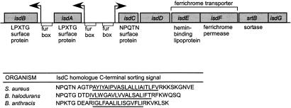
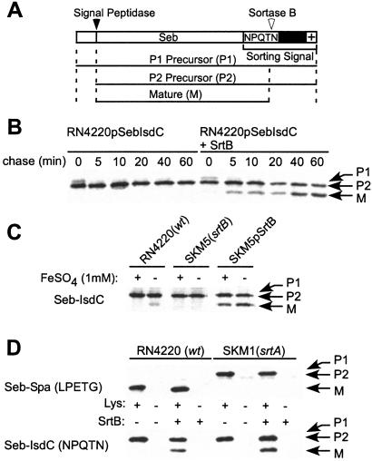
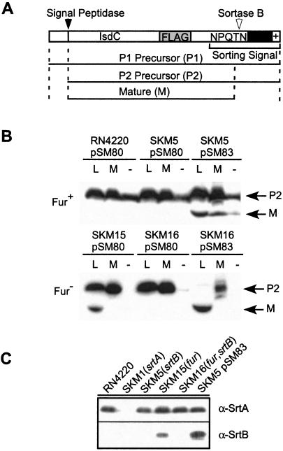
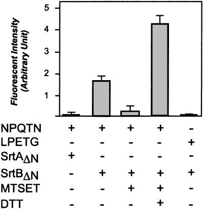
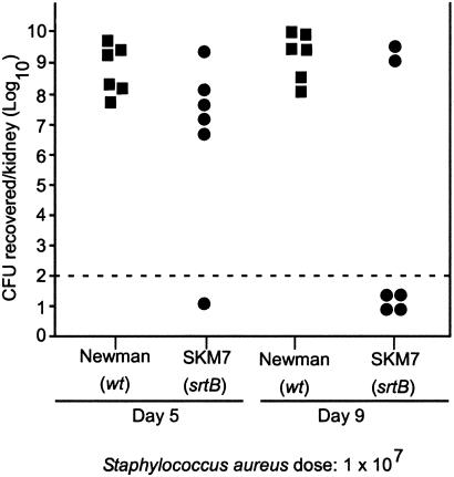
Sortase (SrtA), an enzyme that anchors surface proteins to the cell wall of Gram-positive bacteria, cleaves sorting signals at the LPXTG motif. We have identified a second sortase (SrtB) in the Gram-positive pathogen Staphylococcus aureus that is required for anchoring of a surface protein with a NPQTN motif. Purified SrtB cleaves NPQTN-bearing peptides in vitro, and a srtB mutant is defective in the persistence of animal infections. srtB is part of an iron-regulated locus called iron-responsive surface determinants (isd), which also contains a ferrichrome transporter and surface proteins with NPQTN and LPXTG motifs. Cell wall-anchored surface proteins and the isd locus seem involved in a novel mechanism of iron acquisition that is important for bacterial pathogenesis.


Figures

Figure 1
Figure 1
Genomic organization of S. aureus isd. srtB is located in an operon with a putative iron transporter (isdE, isdF) and a surface protein with a C-terminal NPQTN sorting signal (isdC). Adjacent to this operon are two LPXTG-containing surface proteins that are substrates for srtA-mediated anchoring as shown in Table 2. All transcriptional units contain a Fur box regulatory element upstream of their putative promoters (arrows). The peptide sequence of C-terminal sorting signals of IsdC from S. aureus is compared with that of Bacillus halodurans and Bacillus anthracis.
Figure 2
Figure 2
IsdC contains a C-terminal NPQTN sorting signal and is cleaved by SrtB. (A) The drawing shows the fusion of the C-terminal sorting signal of IsdC to Seb. The cytoplasmic precursor (P1) is cleaved by signal peptidase, generating the extracellular P2 precursor. SrtB cleaves P2 and anchors the mature (M) species to the cell wall. (B) S. aureus RN4220 harboring pSM74 or pSM75 was subjected to pulse labeling with [35S]methionine, and Seb-IsdC was immunoprecipitated and analyzed on SDS/PAGE. pSM74 encodes seb-isdC, whereas pSM75 encodes seb-isdC as well as srtB under control of the srtA promoter. (C) S. aureus RN4220 (pSM74), SKM5 (srtB) (pSM74) and SKM5 (pSM75) were pulse-labeled in the presence or absence of 1 mM FeSO4 and incubated for 30 min. wt, wild type. (D) S. aureus RN4220 and SKM1 (srtA) harboring pSM74, pSM75, pSM79, or pSeb-Spa (Seb with the LPXTG sorting signal or protein A) were pulse-labeled in the presence or absence of 100 mM NH2OH. pSM79 encodes the seb-spa fusion of pSeb-Spa and srtB under control of the srtA promoter. After trichloroacetic acid precipitation, samples were treated with 10 μg/ml lysostaphin or left untreated and then boiled in 4% SDS buffer. Immunoprecipitated Seb-Spa and Seb-IsdC were analyzed by SDS/PAGE and PhosphorImager.
Figure 3
Figure 3
Fur-regulated cell wall anchoring of IsdC requires SrtB. (A) The FLAG epitope tag was introduced into the coding sequence of isdC, and the gene was expressed from the seb promoter in pSM80. The drawing shows the primary structure of IsdCFLAG. The cytoplasmic precursor (P1) is cleaved by signal peptidase, generating extracellular P2. The P2 precursor is cleaved by SrtB, tethering the mature (M) species to the cell wall. (B) After transformation with pSM80 or pSM83, a pSM80 derivative encoding srtB under control of the srtA promoter, S. aureus cultures were precipitated with trichloroacetic acid and treated with the muralytic enzymes lysostaphin (L) and mutanolysin (M) or left untreated (−). Proteins were separated on SDS/PAGE, and IsdCFLAG was identified by immunoblotting with FLAG-specific monoclonal antibody. (C) Immunoblotting of proteins from lysostaphin-treated staphylococcal extracts with specific rabbit antibody reveals the expression of srtA and srtB in various S. aureus strains.
Figure 4
Figure 4
Purified SrtB cleaves NPQTN peptides in vitro. SrtAΔN and SrtBΔN, recombinant sortases with a six-histidine tag replacing the N-terminal membrane anchor, were purified from E. coli extracts by using affinity chromatography. Sortase enzymes were incubated with the LPETG or NPQTN peptides, and cleavage was measured as an increase in fluorescence intensity. [2-(trimethylammonium)ethyl]methanethiosulfonate (MTSET) or DTT were added as indicated.
Figure 5
Figure 5
Mutants in srtB are defective in persistence of renal infections of mice. Swiss–Webster mice were injected intravenously with 1 × 10⁷ colony forming units (cfus) of the S. aureus clinical isolate Newman or the isogenic srtB mutant (SKM7). After 5 or 9 days, animals were killed, their kidneys were harvested, homogenized in 1% Triton-X100, and incubated on agar plates, and staphylococcal colonies were counted. Each symbol (■, wild type; ●, srtB mutant) represents the staphylococcal count in the kidneys of one animal. The dashed line represents the limit of detection for staphylococci in this assay system.

References

    1. Braun V. Science. 1998;282:2202–2203. - PubMed
    1. Weinberg E D. Biometals. 2000;13:85–89. - PubMed
    1. Drazek E S, Hammack C A, Sr, Schmitt M P. Mol Microbiol. 2000;36:68–84. - PubMed
    1. Hantke K. Curr Opin Microbiol. 2001;4:172–177. - PubMed
    1. Schmitt M P. J Bacteriol. 1997;179:838–845. - PMC - PubMed

Publication types

LinkOut - more resources