. 2002 Feb 19;99(4):1853-8.
doi: 10.1073/pnas.032613199. Epub 2002 Feb 5.

A poxvirus-like type IB topoisomerase family in bacteria

Affiliations

A poxvirus-like type IB topoisomerase family in bacteria

Berit Olsen Krogh et al. Proc Natl Acad Sci U S A. .

Abstract

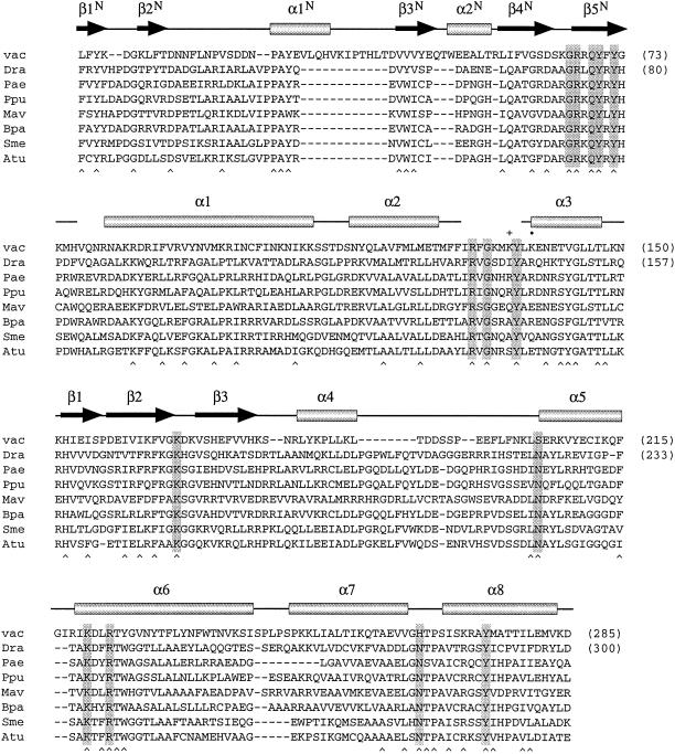
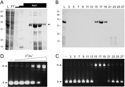
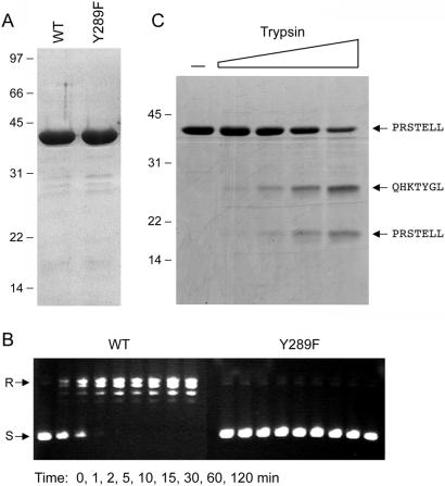
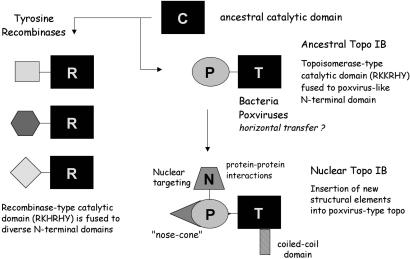
We report that diverse species of bacteria encode a type IB DNA topoisomerase that resembles vaccinia virus topoisomerase. Deinococcus radiodurans topoisomerase IB (DraTopIB), an exemplary member of this family, relaxes supercoiled DNA in the absence of a divalent cation or ATP. DraTopIB has a compact size (346 aa) and is a monomer in solution. Mutational analysis shows that the active site of DraTopIB is composed of the same constellation of catalytic side chains as the vaccinia enzyme. Sequence comparisons and limited proteolysis suggest that their folds are conserved. These findings imply an intimate evolutionary relationship between the poxvirus and bacterial type IB enzymes, and they engender a scheme for the evolution of topoisomerase IB and tyrosine recombinases from a common ancestral strand transferase in the bacterial domain. Remarkably, bacteria that possess topoisomerase IB appear to lack DNA topoisomerase III.


Figures

Figure 1
Figure 1
A family of bacterial type IB topoisomerases related to vaccinia virus topoisomerase. The amino acid sequence of vaccinia virus topoisomerase (vac) from positions 4 to 285 is aligned with the sequences of putative topoisomerases encoded by Deinococcus radiodurans (Dra), Pseudomonas aeruginosa (Pae), Pseudomonas putida (Ppu), Mycobacterium avium (Mav), Bordetella parapertussis (Bpa), Sinorhizobium meliloti (Sme), and Agrobacterium tumefaciens (Atu). The secondary structural elements of vaccinia topoisomerase are shown above its sequence. Conserved residues involved in DNA binding or transesterification by vaccinia topoisomerase are highlighted in shaded boxes. Positions of side-chain identity or similarity in all eight of the proteins are indicated by a ∧ below the sequences.
Figure 2
Figure 2
Purification and supercoil relaxation activity of DraTopIB. (A) Aliquots (15 μl) of the soluble bacterial lysate (L), the phosphocellulose flow-through (FT), the 0.1 and 0.35 M NaCl washes (first two NaCl “steps”), and sequential fractions from the 1 M NaCl elution (top NaCl step) were analyzed by SDS/PAGE. The gel was stained with Coomassie blue dye. The positions and sizes (in kDa) of marker polypeptides are indicated on the left. The DraTopIB polypeptide is denoted by the arrow at right. (B) Glycerol gradient sedimentation. Aliquots (20 μl) of the odd-numbered gradient fractions were analyzed by SDS/PAGE. An aliquot of the phosphocellulose load fraction is included in lane L. (C) DNA relaxation reaction mixtures containing 50 mM Tris⋅HCl (pH 7.5), 2.5 mM EDTA, 100 mM NaCl, 0.3 μg of supercoiled pUC19 DNA, and 0.5 μl of the indicated gradient fractions were incubated for 2 h at 37°C. The DNA products were analyzed by agarose gel electrophoresis. A photograph of the ethidium bromide-stained gel is shown; the positions of supercoiled (S) and relaxed (R) circular DNAs are indicated on the left. (D) Stimulation of relaxation by potassium glutamate. Reaction mixtures containing 50 mM potassium phosphate buffer (pH 8.0), 0.3 μg of pUC19 DNA, 350 ng of DraTopIB, and 5, 10, 20, 50, 100, or 200 mM potassium glutamate were incubated for 15 min at 37°C. DraTopIB was omitted from the control reaction in lane −.
Figure 3
Figure 3
Probing the mechanism and structure of DraTopIB. (A) Aliquots (5 μg) of wild-type DraTopIB and the Y289F mutant were analyzed by SDS/PAGE. A Coomassie blue-stained gel is shown. (B) Relaxation reaction mixtures containing (per 20 μl) 50 mM potassium phosphate (pH 8.0), 200 mM potassium glutamate, 0.3 μg of pUC19, and 350 ng of enzyme were incubated at 37°C. Aliquots (20 μl) were withdrawn at the times specified and quenched immediately with SDS. The DNA products were analyzed by agarose gel electrophoresis. (C) Mixtures (20 μl) containing 50 mM Tris⋅HCl (pH 7.5), 4 μg of wild-type DraTopIB, and 0, 10, 20, 40, or 80 ng of trypsin were incubated on ice for 15 min. The digestion products were resolved by SDS/PAGE. The gel contents were electroblotted to a poly(vinylidene difluoride) membrane. A picture of the Coomassie blue-stained membrane is shown. The amino-terminal amino acid sequences of intact DraTopIB and the two major tryptic fragments were determined by automated Edman chemistry and are indicated on the right.
Figure 4
Figure 4
Defining the active site of DraTopIB. (A) The indicated DraTopIB proteins (2 μg) were analyzed by SDS/PAGE. A Coomassie blue-stained gel is shown. (B and C) Relaxation reaction mixtures containing (per 20 μl) 50 mM potassium phosphate (pH 8.0), 200 mM potassium glutamate, 0.3 μg of pUC19 DNA, and 350 ng of enzyme were incubated at 37°C. Aliquots (20 μl) were withdrawn at the times specified and quenched immediately with SDS. One-half of each sample was analyzed by agarose gel electrophoresis, after which the gels were stained with ethidium bromide (B). The other one-half of each sample was electrophoresed through an agarose gel containing 0.5 μg/ml ethidium bromide (C). The positions of supercoiled (S), relaxed (R), and nicked (N) circular DNAs are indicated on the right.
Figure 5
Figure 5
Evolution of the topo IB family. See text for details.

References

    1. Shuman S. Biochim Biophys Acta. 1998;1400:321–337. - PubMed
    1. Champoux J J. Annu Rev Biochem. 2001;70:369–413. - PubMed
    1. Petersen B Ø, Shuman S. J Biol Chem. 1997;272:3891–3896. - PubMed
    1. Wittschieben J, Shuman S. Nucleic Acids Res. 1997;25:3001–3008. - PMC - PubMed
    1. Cheng C, Wang L K, Sekiguchi J, Shuman S. J Biol Chem. 1997;272:8263–8269. - PubMed

Publication types

MeSH terms