. 2002 Feb 19;99(4):1899-904.
doi: 10.1073/pnas.042529199. Epub 2002 Feb 5.

Regulation of the tyrosine kinase Itk by the peptidyl-prolyl isomerase cyclophilin A

Affiliations

Regulation of the tyrosine kinase Itk by the peptidyl-prolyl isomerase cyclophilin A

Kristine N Brazin et al. Proc Natl Acad Sci U S A. .

Abstract

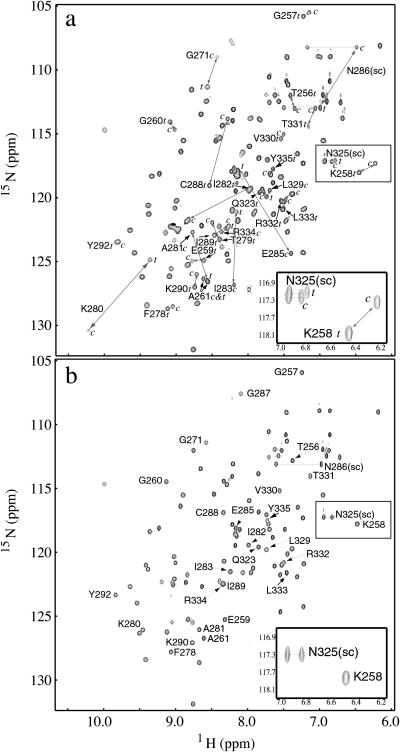
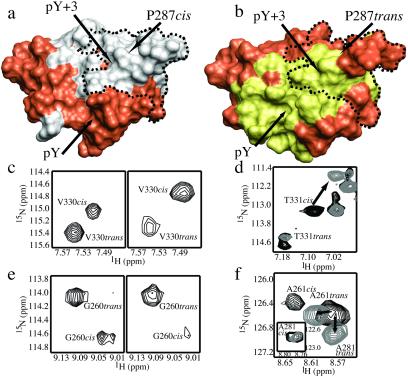
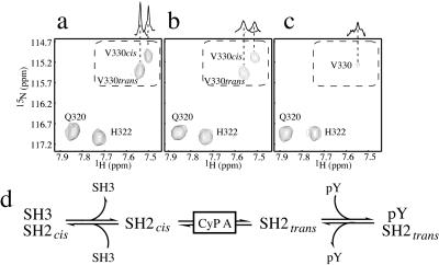
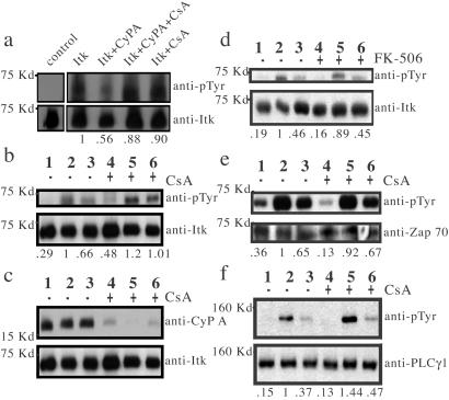
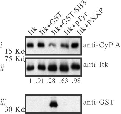
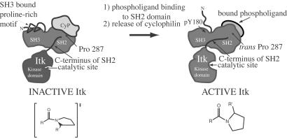
Interleukin-2 tyrosine kinase (Itk) is a nonreceptor protein tyrosine kinase of the Tec family that participates in the intracellular signaling events leading to T cell activation. Tec family members contain the conserved SH3, SH2, and catalytic domains common to many kinase families, but they are distinguished by unique sequences outside of this region. The mechanism by which Itk and related Tec kinases are regulated is not well understood. Our studies indicate that Itk catalytic activity is inhibited by the peptidyl prolyl isomerase activity of cyclophilin A (CypA). NMR structural studies combined with mutational analysis show that a proline-dependent conformational switch within the Itk SH2 domain regulates substrate recognition and mediates regulatory interactions with the active site of CypA. CypA and Itk form a stable complex in Jurkat T cells that is disrupted by treatment with cyclosporin A. Moreover, the phosphorylation levels of Itk and a downstream substrate of Itk, PLCgamma1, are increased in Jurkat T cells that have been treated with cyclosporin A. These findings support a novel mode of tyrosine kinase regulation for a Tec family member and provide a molecular basis for understanding a cellular function of the ubiquitous peptidyl prolyl isomerase, CypA.


Figures

Figure 1
Figure 1
The Itk SH2 domain adopts two stable structures in solution. (a) 1H-15N HSQC spectrum of purified, recombinant 15N-labeled Itk SH2 domain. The resonances corresponding to the cis (c) and trans (t) conformers are connected by a gray double-headed arrow. (Inset) An expansion of the boxed region of the spectrum containing representative doubled resonances for the Asn-325 side chain (sc) NH2 and the Lys-258 backbone NH. (b) HSQC spectrum of the Itk SH2 domain in which Pro-287 is mutated to Gly. Labeled cross-peaks correspond to those that are labeled in a, and the cross-peak corresponding to Gly-287 (introduced by mutation) is assigned. (Inset) The same region as in a but showing a single resonance for Asn-325 (sc) and Lys-258.
Figure 2
Figure 2
Surface representations (rendered with molmol; ref. 51) of the Itk SH2 cis (a) and trans (b) structural models (R.J.M. and A.H.A., unpublished results). The SH2 residues that display conformational heterogeneity are demarcated with a dotted line on each model. Pro-287 is indicated, and the pY and pY + 3 binding pockets are labeled. Chemical shift perturbations that occur on addition of equimolar Itk SH3 domain to the Itk SH2 domain (38) are mapped onto the cis model and highlighted in gray (a). Likewise, SH2 residues that undergo chemical shift perturbations on addition of three times excess phosphopeptide (ADpYEPPPSNDE) are highlighted in yellow on the trans model (b). For both surface models, residues that do not exhibit chemical shift changes on addition of ligand are orange. (c) Select region of the HSQC spectrum of a 0.5 mM Itk SH2 sample. (Left) Itk SH2 domain alone, where the trans/cis ratio is approximately 60:40 based on the cross-peak volumes for each set of doubled resonances. (Right) The same region of the Itk SH2 HSQC spectrum after addition of 10 times excess unlabeled Itk SH3 domain (trans/cis ratio 10:90). (d) Superposition of the same region of two HSQC spectra of 15N-labeled Itk SH2 domain in the absence (black) and presence (gray) of unlabeled Itk SH3 domain. The Thr-331 cis cross-peak shifts on addition of equimolar Itk SH3 domain (arrow), whereas the Thr-331 trans cross-peak remains at the same frequency. (e) (Left) Itk SH2 domain (trans/cis 60:40). (Right) Addition of 3 times excess phosphopeptide to Itk SH2 (trans/cis 90:10). (f) Superposition of two HSQC spectra of 15N-labeled Itk SH2 domain in the absence (black) and presence (gray) of 3 times excess phosphopeptide. The Ala-261 and Ala-281 trans cross-peaks shift on addition of phosphopeptide (arrows), whereas the Ala-261 and Ala-281 cis cross-peaks resonate at the same frequency regardless of ligand present (the Ala-281 cis cross-peak is shown in the Inset because of the large difference in resonance frequencies for the cis and trans resonances of Ala-281).
Figure 3
Figure 3
(a) Region of the HSQC spectrum for the 15N-labeled Itk SH2 domain (0.5 mM) that includes cross-peaks for the Gln-320, His-322, and Val-330 backbone NH resonances. Gln-320 and His-322 are not affected by cis/trans isomerization around the Asn-286–Pro-287 imide bond; therefore, each appears as a single cross-peak. In contrast, the Val-330 amide resonance is doubled as a result of slow exchange between the cis and trans forms. (b) Same region of the HSQC spectrum for the Itk SH2 domain (0.5 mM) in the presence of 3 mol% CypA. Line broadening is apparent for all of the doubled resonances in the SH2 domain spectrum on addition of CypA, whereas those peaks that correspond to residues unaffected by the isomerization event do not broaden significantly. (c) Addition of 20 mol% CypA to the Itk SH2 domain results in further line broadening and coalescence to the chemical shift value that represents the average of the cis and trans conformers. One-dimensional projections through the center of the Val-330 cross-peaks along the 1H axis illustrate the exchange mediated broadening and coalescence. (d) Equilibrium model encompassing the CypA-catalyzed Itk SH2 cis/trans interconversion, cis-mediated SH3 binding (Itk dimerization), and trans-mediated phospholigand (pY) binding.
Figure 4
Figure 4
(a) CypA, CypA/CsA complex, or CsA was added to baculovirus-expressed Itk immediately before resuspension in ATP/kinase buffer. A mock kinase assay of Itk (left lane) was performed in which ATP was excluded from the kinase reaction buffer. Itk phosphorylation is normalized to Itk protein levels, and net changes in phosphorylation are indicated below each lane. (b–f) Lanes 1, 2, and 3 are mock stimulation, 40 s and 2 min, respectively, following TCR stimulation of Jurkat cells in the absence (−) of drug treatment, whereas lanes 4, 5, and 6 represent the same conditions in the presence (+) of drug. (b) Itk phosphorylation following TCR stimulation of Jurkat cells in the presence and absence of CsA. (c) CypA binding to Itk was monitored by detection of Itk immunoprecipitates with an anti-CypA antibody. (d) Itk phosphorylation in the presence and absence of FK-506. (e) Zap-70 phosphorylation and (f) PLCγ1 phosphorylation in the presence and absence of CsA. Phosphorylation levels of Itk, Zap-70, and PLCγ1 are normalized to total protein in each lane. Net changes in phosphorylation relative to phosphorylation levels at 40 s following stimulation in the absence of drug (lane 2) are indicated below each lane.
Figure 5
Figure 5
For the indicated samples, glutathione S-transferase (GST), Itk GST-SH3, phosphotyrosine-containing peptide (pTyr), or a proline-rich peptide (PXXP) was incubated with full-length Itk following immunoprecipitation from Jurkat T-cell lysates. (Top, i) Following extensive washing, the amount of CypA that coimmunoprecipitated with Itk was determined by immunoblotting. The amount of CypA in Itk immunoprecipitates is normalized to total Itk in each lane. Net changes in the amount of coimmunoprecipitated CypA relative to the amount that coimmunoprecipitates with Itk in the absence of exogenous factors are indicated. (Bottom, iii) Detection of GST-SH3 in Itk immunoprecipitates following competitive binding assay.
Figure 6
Figure 6
Model for Itk regulation. Itk is dark gray and includes the proline-rich region, SH3, SH2, and kinase domains (the PH domain and part of the TH domain are not shown for clarity). Two “states” of Itk are depicted: inactive, CypA-bound Itk; and active, phospholigand-bound Itk. CypA is light gray and is shown bound to the Itk SH2 domain in the region of Pro-287. The proposed configuration of the Asn-286–Pro-287 imide bond (resembling the transition state for cis/trans interconversion) is illustrated for the inactive, CypA-bound form of Itk. NMR spectroscopic data suggest that cyclophilin-bound Itk is monomeric (K.N.B. and A.H.A., unpublished results), and the proline-rich region may contact the SH3 binding pocket in an intramolecular fashion (52). Activation may be accompanied by phosphorylation of Tyr-180 in the SH3 domain expelling bound proline (53). Cis/trans isomerization around Asn-286–Pro-287 causes pronounced conformational heterogeneity in the C terminus of the Itk SH2 domain leading directly into the Itk kinase domain (see Fig. 1). The C terminus of the Itk SH2 domain may adopt a conformation in the presence of cyclophilin that structurally perturbs the kinase catalytic site rendering it inactive. Release of cyclophilin, either by Itk dimerization to favor the cis conformer (not shown) or by binding of a phosphotyrosine-containing ligand to favor the trans conformer, may lead to reorganization of the C-terminal SH2 residues and subsequent restructuring of the kinase active site.

Similar articles

Cited by

References

    1. Hubbard S R, Till J H. Annu Rev Biochem. 2000;69:373–398. - PubMed
    1. Xu W, Harrison S C, Eck M J. Nature (London) 1997;385:595–602. - PubMed
    1. Sicheri F, Moarefi I, Kuriyan J. Nature (London) 1997;385:602–609. - PubMed
    1. Willams J C, Wierenga R K, Saraste M. Trends Biochem Sci. 1998;23:179–184. - PubMed
    1. Yang W-C, Collette Y, Nunès J A, Olive D. Immunity. 2000;12:373–382. - PubMed

Publication types

MeSH terms

LinkOut - more resources