. 2002 Feb 19;99(4):1943-8.
doi: 10.1073/pnas.042688399. Epub 2002 Feb 5.

Identification of a functional role for lipid asymmetry in biological membranes: Phosphatidylserine-skeletal protein interactions modulate membrane stability

Affiliations

Identification of a functional role for lipid asymmetry in biological membranes: Phosphatidylserine-skeletal protein interactions modulate membrane stability

Sumie Manno et al. Proc Natl Acad Sci U S A. .

Abstract

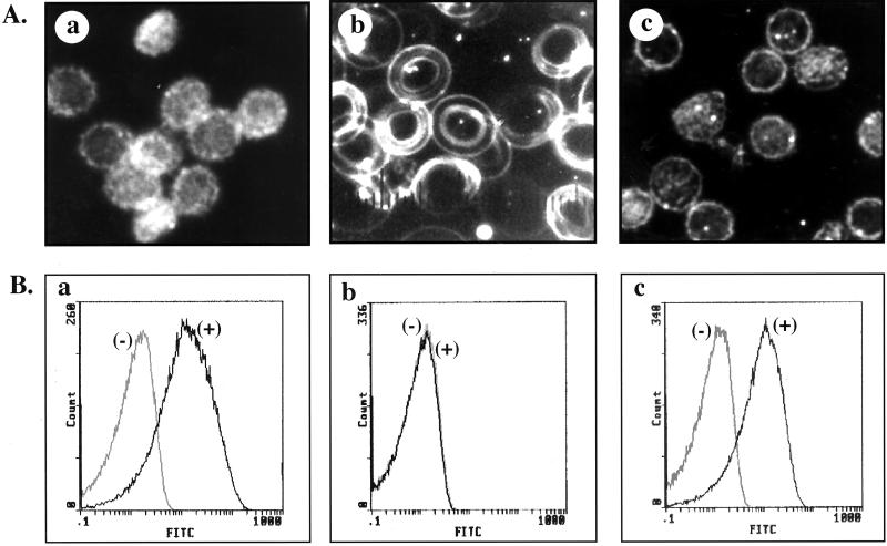
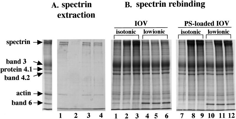
Asymmetric distribution of phospholipids is ubiquitous in the plasma membranes of many eukaryotic cells. The majority of the aminophospholipids are located in the inner leaflet whereas the cholinephospholipids are localized predominantly in the outer leaflet. Several functional roles for asymmetric phospholipid distribution in plasma membranes have been suggested. Disruption of lipid asymmetry creates a procoagulant surface on platelets and serves as a trigger for macrophage recognition of apoptotic cells. Furthermore, the dynamic process of phospholipid translocation regulates important cellular events such as membrane budding and endocytosis. In the present study, we used the red cell membrane as the model system to explore the contribution of phospholipid asymmetry to the maintenance of membrane mechanical properties. We prepared two different types of membranes in terms of their phospholipid distribution, one in which phospholipids were scrambled and the other in which the asymmetric distribution of phospholipids was maintained and quantitated their mechanical properties. We documented that maintenance of asymmetric distribution of phospholipids resulted in improved membrane mechanical stability. The greater difficulty in extracting the spectrin-actin complex at low-ionic strength from the membranes with asymmetric phospholipid distribution further suggested the involvement of interactions between aminophospholipids in the inner leaflet and skeletal proteins in modulating mechanical stability of the red cell membrane. These findings have enabled us to document a functional role of lipid asymmetry in regulating membrane material properties.


Figures

Figure 1
Figure 1
The morphology and the binding of FITC-labeled annexin V to various resealed membrane preparations. (A) Dark-field light microscopy of control ghosts (a), MgATP-ghosts (b), and MgAMPPNP-ghosts (c). Control and MgAMPPNP-ghosts are echinocytic whereas MgATP ghosts are discocytic. (B) Representative cytofluorograph histograms of various resealed membrane preparations after binding of FITC-labeled annexin V. Histograms in gray represent background fluorescence whereas histograms in black represent specific binding. Control ghosts (a) and MgAMPPNP-ghosts (c) exhibit significant binding of annexin V whereas there is no binding of annexin V to MgATP-ghosts (b).
Figure 2
Figure 2
Membrane mechanical properties of various resealed membrane preparations. Membrane deformability (A) and mechanical stability (B) were measured by using an ektacytometer. (A) The membrane deformability profile of MgATP-ghosts is virtually the same as that of control-ghosts and MgAMPPNP-ghosts. (B) The rate of decrease in deformability index of MgATP-ghosts was slower than that for control ghosts, implying increased membrane mechanical stability of MgATP ghosts. In marked contrast, fragmentation pattern of MgAMPPNP-ghosts was the same as that of control ghosts.
Figure 3
Figure 3
Effects of various ATPase inhibitors and of different MgATP concentrations on membrane mechanical stability of MgATP-ghosts. (A) The rate of decrease in the deformability index of MgATP-ghosts treated with either Ouabain (10 μM) or EGTA (0.1 mM) was the same as MgATP ghosts, implying that these two ATPase inhibitors had no effect on mechanical stability. In marked contrast, MgATP-ghosts treated with either Vanadate (10 μM) or PDA (10 mM) fragmented at the same rate as control ghosts and at a much faster rate than untreated MgATP ghosts, implying that these two ATPase inhibitors abolished the effect of MgATP on membrane mechanical stability. (B) Effect of various MgATP concentrations (0.0 to 0.8 mM) on membrane mechanical stability. The rate of decrease in the deformability index decreased with increasing concentrations of MgATP.
Figure 4
Figure 4
Interaction of spectrin with membranes in various ghost preparations (A) Protein composition of low ionic buffer (0.5 mM phosphate buffer, pH 8.0) extracts from control ghosts (lane 1), MgATP-ghosts (lane 2), MgATP-ghosts with vanadate (lane 3), and MgAMPPNP-ghosts (lane 4). The far left lane shows the protein composition of native red cell membranes. Note spectrin and actin are extracted from the membranes of control, MgATP ghosts treated with vanadate and MgAMPPNP ghosts but not from MgATP ghosts. (B) Rebinding of purified spectrin dimer to IOVs and PS-loaded IOVs. Spectrin dimer at concentrations of 0.75 mg/ml (lanes 2, 5, 8, and 11) or 1.13 mg/ml (lanes 3, 6, 9, and 12) was added to 0.5 mg of IOVs or PS-loaded IOVs. Spectrin bound to IOVs in isotonic buffer (lanes 2 and 3) but not in low ionic buffer (lanes 5 and 6). However, spectrin bound to PS-loaded IOVs under both isotonic (lanes 8 and 9) and low ionic (lanes 11 and 12) conditions. The protein composition of native IOVs (lanes 1 and 4) and PS-loaded IOVs (lanes 7 and 10) before addition of spectrin are also shown. Note that band 6 remains bound to IOVs under low ionic conditions but is released from IOVs under isotonic conditions.

References

    1. Devaux PF. Biochemistry. 1991;30:1163–1173. - PubMed
    1. Williamson P, Schlegel R A. Mol Membr Biol. 1994;11:199–216. - PubMed
    1. Tang X, Halleck M S, Schlegel R A, Williamson P. Science. 1996;272:1495–1497. - PubMed
    1. Devaux PF. Annu Rev Biophys Biomol Struct. 1992;21:417–439. - PubMed
    1. Zwaal R F A, Schroit A J. Blood. 1997;89:1121–1132. - PubMed

Publication types

LinkOut - more resources