. 2002 Feb 5;99(3):1627-32.
doi: 10.1073/pnas.251673298.

The SNARE protein SNAP-25 is linked to fast calcium triggering of exocytosis

Affiliations

The SNARE protein SNAP-25 is linked to fast calcium triggering of exocytosis

Jakob B Sørensen et al. Proc Natl Acad Sci U S A. .

Erratum in

  • Proc Natl Acad Sci U S A 2002 Apr 30;99(9):6449

Abstract

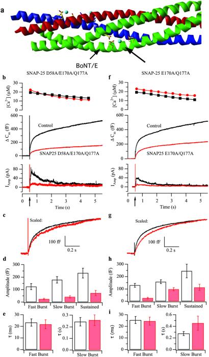
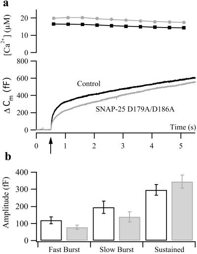
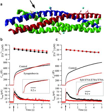
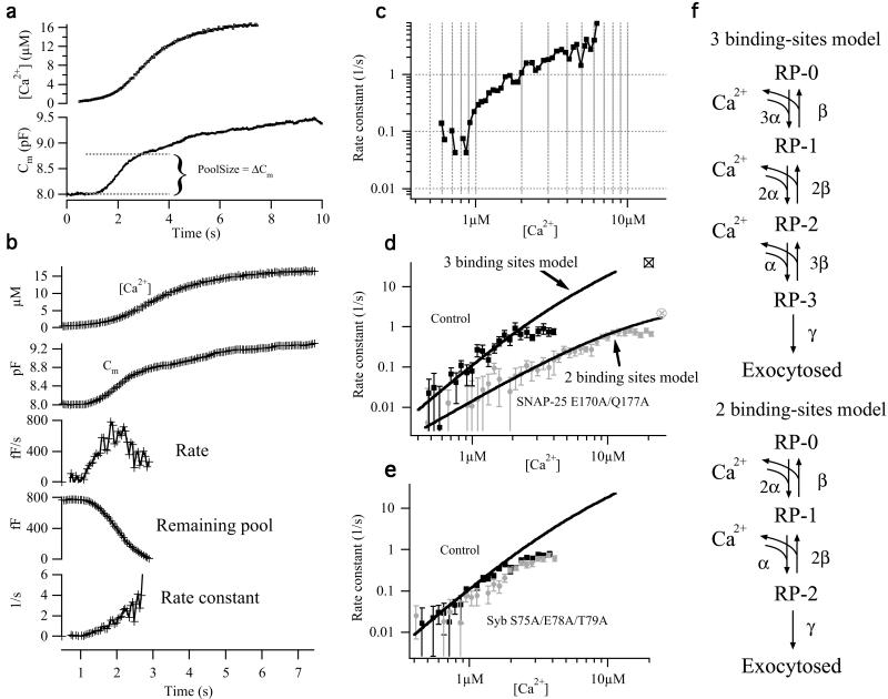
Synchronous neurotransmission depends on the tight coupling between Ca(2+) influx and fusion of neurotransmitter-filled vesicles with the plasma membrane. The vesicular soluble N-ethylmaleimide-sensitive factor attachment protein receptor (SNARE) protein synaptobrevin 2 and the plasma membrane SNAREs syntaxin 1 and synaptosomal protein of 25 kDa (SNAP-25) are essential for calcium-triggered exocytosis. However, the link between calcium triggering and SNARE function remains elusive. Here we describe mutations in two sites on the surface of the SNARE complex formed by acidic and hydrophilic residues of SNAP-25 and synaptobrevin 2, which were found to coordinate divalent cations in the neuronal SNARE complex crystal structure. By reducing the net charge of the site in SNAP-25 we identify a mutation that interferes with calcium triggering of exocytosis when overexpressed in chromaffin cells. Exocytosis was elicited by photorelease of calcium from a calcium cage and evaluated by using patch-clamp capacitance measurements at millisecond time resolution. We present a method for monitoring the dependence of exocytotic rate upon calcium concentration at the release site and demonstrate that the mutation decreased the steepness of this relationship, indicating that the number of sequential calcium-binding steps preceding exocytosis is reduced by one. We conclude that the SNARE complex is linked directly to calcium triggering of exocytosis, most likely in a complex with auxiliary proteins.


Figures

Figure 1
Figure 1
Mutation in the site D58/E170/Q177 in SNAP-25 inhibits secretion. (a) Crystal structure of the core complex. Red, syntaxin 1; blue, synaptobrevin 2; green, SNAP-25; and light blue, strontium ions (16). Membrane anchors (not shown) link to the complex at the left side. Side chains of amino acids interacting with strontium ions are shown as sticks. The arrows point to the site studied in this figure and the botulinum toxin E (BoNT/E) cleavage site. (b) Secretion from chromaffin cells after flash photorelease of calcium. Shown are means of 31 control (not expressing) cells (black) and 29 cells overexpressing SNAP-25 D58A/E170A/Q177A (red). (Top) [Ca2+]i after flash photolysis of nitrophenyl-EGTA (arrow on the time scale). Preflash [Ca2+]i was 200–500 nM. (Middle) Mean capacitance change. (Bottom) Mean amperometric current. (c) Mean capacitance change from mutant cells (red) scaled to the amplitude of control data (black) at 1 s after the flash. (d and e) Amplitudes and time constants (mean ± SEM) of exponential fits to individual responses. (f–i) Double mutation in the site D58/E170/Q177 in SNAP-25 slows down secretion. Means of 47 control cells (black) and 36 cells overexpressing SNAP-25 E170A/Q177A (red) are shown. For explanation, see b–e legends.
Figure 2
Figure 2
The double charge-neutralizing mutation D179A/186A in the C-terminal end of SNAP-25 moderately decreases secretion. The means of 33 control cells (black) and 30 cells overexpressing SNAP-25 D179A/186A are shown (gray). For explanation, see Fig. 1 b and d legends.
Figure 3
Figure 3
Triple mutation in the site S75/E78/T79 in synaptobrevin blocks the burst phase of secretion. (a) Crystal structure of the core complex. Red, syntaxin 1; blue, synaptobrevin 2; green, SNAP-25; and light blue, strontium ions. The arrow points to the studied site. (b) The means of 51 control (not expressing) cells (black) and 35 cells overexpressing synaptobrevin wild type (red). (Top) Mean [Ca2+]i after the flash. (Middle) Mean capacitance change. (Bottom) Mean amperometric current. (Inset) The mean trace from cells overexpressing synaptobrevin wild type scaled to the control data at 1 s after the flash. (c) The means of 47 control cells (black) and 42 cells overexpressing synaptobrevin S75A/E78A/T79A (red). Explanation is the same as that for b.
Figure 4
Figure 4
Double mutation in the D58/E170/Q177 site changes the calcium dependence of exocytosis. (a) Example of a control cell stimulated by continuous weak UV illumination. Capacitance (line, Bottom) and calcium (small boxes, Top) measurements are shown. (b) Rate constant calculations. The capacitance trace was averaged (second trace) around each calcium measurement (first trace), and the local rate was calculated (third trace). Based on an estimate of the size of the readily releasable pool (see a), the remaining pool size was calculated (fourth trace), and finally the rate constant (fifth trace) was calculated as the rate divided by remaining pool size. (c) Plot of rate constant versus [Ca2+]i on a double logarithmic scale. (d) Double logarithmic plot of rate constant versus calcium for 15 control cells (black squares) and 13 cells overexpressing the SNAP-25 E170A/Q177A mutant (gray circles). For comparison with ramp data, the rates for the corresponding flash data (from fig. 1i) are indicated with open symbols with crosses. For the control cells the rate for the readily releasable pool was taken, and for the mutant cells the rate for the slowly releasable pool was chosen. The flash rates confirm that the apparent saturation of ramp rates at around 1 s is artificial, probably caused by some contamination of the pool size estimate with slower components at later times. Fitted to control data are the three binding-sites secretory model in f, whereas fit to mutant data are the two binding-sites model. (e) Double logarithmic plot of rate constant versus calcium for 13 control cells (black squares) and 13 cells overexpressing the synaptobrevin S75A/E78A/T79A mutant (gray circles). The line is the same fit of the three-step model as the one in a. (f) Sequential calcium-binding models for exocytosis. (Upper) Standard model for chromaffin cells, with three equivalent calcium-binding sites. (Lower) Modified model, with only two equivalent calcium-binding sites. The models were solved numerically by using a fourth order Runge-Kutta method with automatic step-size control and a typical calcium ramp as forcing function. A simplex method was used to fit the models to the estimated data in d. Fitted parameters in the three binding-sites model (fit to controls in d; only α was varied, β and γ were taken from ref. 12): α = 5.7 μM−1⋅s−1; β = 56 s−1; and γ = 1450 s−1. Fitted parameters in the two binding-sites model (fit to SNAP-25 E170/177A data): α = 4.2 μM−1⋅s−1; β = 69 s−1; and γ = 4.7 s−1.

References

    1. Söllner T, Whitehart S W, Brunner M, Erdjument-Bromage H, Geromanos S, Tempst P, Rothman J E. Nature (London) 1993;362:318–324. - PubMed
    1. Jahn J, Südhof T C. Annu Rev Biochem. 1999;68:863–911. - PubMed
    1. Lin R C, Scheller R H. Annu Rev Cell Dev Biol. 2000;16:19–49. - PubMed
    1. Brose N, Petrenko A G, Südhof T C, Jahn R. Science. 1992;256:1021–1025. - PubMed
    1. Fernandez-Chacon R, Königstorfer A, Gerber S H, Garcia J, Matos M F, Stevens C F, Brose N, Rizo J, Rosenmund C, Südhof T C. Nature (London) 2001;410:41–49. - PubMed

Publication types

MeSH terms

LinkOut - more resources