

Experimental models to study cholangiocyte biology
- PMID: 11833061
- PMCID: PMC4656596
- DOI: 10.3748/wjg.v8.i1.1
Experimental models to study cholangiocyte biology
Abstract
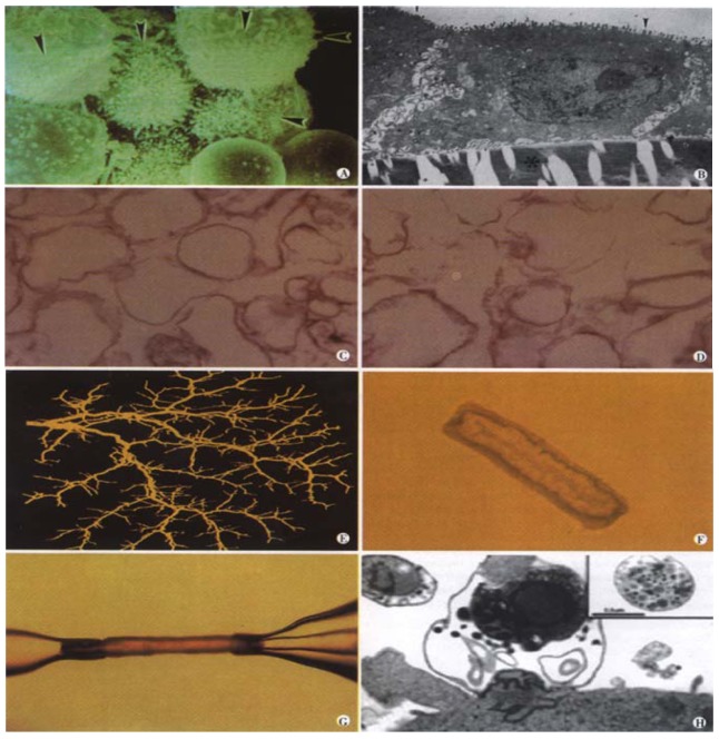
Cholangiocytes-the epithelial cells which line the bile ducts-are increasingly recognized as important transporting epithelia actively involved in the absorption and secretion of water, ions, and solutes. This recognition is due in part to the recent development of new experimental models. New biologic concepts have emerged including the identification and topography of receptors and flux proteins on the apical and/or basolateral membrane which are involved in the molecular mechanisms of ductal bile secretion. Individually isolated and/or perfused bile duct units from livers of rats and mice serve as new,physiologically relevant in vitro models to study cholangiocyte transport. Biliary tree dimensions and novel insights into anatomic remodeling of proliferating bile ducts have emerged from three-dimensional reconstruction using CT scanning and sophisticated software. Moreover, new pathologic concepts have arisen regarding the interaction of cholangiocytes with pathogens such as Cryptosporidium parvum. These concepts and associated methodologies may provide the framework to develop new therapies for the cholangiopathies, a group of important hepatobiliary diseases in which cholangiocytes are the target cell.
Figures
References
-
- Alpini G, Phillips JO, LaRusso NF. The biology of biliary epithelia.Edited by Arias IM, Boyer JL, Fausto N, Jakoby WB, Schachter DA, Shafritz DA. The Liver: Biology and Pathobiology. New York: Raven Press. 1994:623–653.
-
- Lesage G, Glaser SS, Gubba S, Robertson WE, Phinizy JL, Lasater J, Rodgers RE, Alpini G. Regrowth of the rat biliary tree after 70% partial hepatectomy is coupled to increased secretin-induced ductal secretion. Gastroenterology. 1996;111:1633–1644. - PubMed
-
- LeSage GD, Benedetti A, Glaser S, Marucci L, Tretjak Z, Caligiuri A, Rodgers R, Phinizy JL, Baiocchi L, Francis H, et al. Acute carbon tetrachloride feeding selectively damages large, but not small, cholangiocytes from normal rat liver. Hepatology. 1999;29:307–319. - PubMed
-
- Kossor DC, Goldstein RS, Ngo W, DeNicola DB, Leonard TB, Dulik DM, Meunier PC. Biliary epithelial cell proliferation following alpha-naphthylisothiocyanate (ANIT) treatment: relationship to bile duct obstruction. Fundam Appl Toxicol. 1995;26:51–62. - PubMed
Publication types
MeSH terms
Grants and funding
LinkOut - more resources
Full Text Sources
