. 2002 Jan;200(Pt 1):105-11.
doi: 10.1046/j.0021-8782.2001.00006.x.

Optical anisotropy of a pig tendon under compression

Affiliations

Optical anisotropy of a pig tendon under compression

VeraLúciaC Feitosa et al. J Anat. 2002 Jan.

Abstract

The proximal region of the superficial digital flexor tendon of pigs passes under the tibiotarsal joint, where it is subjected to compressional and tensional forces. This region was divided into a surface portion (sp), which is in direct contact with the bone and into a deep portion (dp), which is the layer opposite the articulating surface. The purpose of this work was to analyse the distribution and organisation of the collagen bundles and proteoglycans in the extracellular matrix in sp and dp. Toluidine-blue-stained sections were analysed under a polarising microscope. Strong basophilia and metachromasia were observed in sp, demonstrating accumulation of proteoglycan in a region bearing compression, but the intensity was reduced the further layers were from the bone. Linear dichroism confirmed that the glycosaminoglycan molecules were disposed predominantly parallel to the longest axis of the collagen fibrils. Birefringence analysis showed a higher molecular order and aggregation of the collagen bundles in areas where the tension was more prominent. The crimp pattern was more regular in dp than in sp, probably as a requirement for tendon stretching. The optical anisotropy exhibited by the collagen bundles also confirmed the helical organisation of the collagen bundles in the tendon. Hyaluronidase digestion caused a decrease in the basophilia, but this was not eliminated, supporting the idea that in the matrix, proteoglycans are not completely available to the enzyme action.


Figures

Fig. 1
Fig. 1
Dorsal view of the superficial digital flexor tendon (SDFT). (a) The SDFT was divided into a proximal region (p), which is under the tibiotarsal joint and is subject to compression in addition to tension, an intermediate region (i) where only tension forces are present, and a distal region (d) region, which also withstands compressive force. (b) A diagrammatic representation of a lateral view of the tendon, showing the sp portion, which remains in contact with the bones of the tibiotarsal joint, and the dp portion, the layer opposite the articulating layer.
Fig. 2
Fig. 2
Linear dichroism of the toluidine-blue-stained sections. (a) and (b) correspond to the absorbance when the longest axis of the tendon is perpendicular and parallel, respectively, to the azimuth of the polariser. Comparison of images (a) with (b) reveals higher absorption when the fibres are perpendicular to the plane of the polarised light. Note the stronger metachromasy in sp, which is near the bone. In the deep layer (dp), the staining and even metachromasy are noticeably reduced. (b) Same section, but with fibres positioned parallel to the polarised light plane. Scale bars = 238 µm.
Fig. 3
Fig. 3
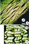
Birefringent images of sections of the proximal region of the SDFT, stained by toluidine blue, pH 4.0. The main axis of the tendon was positioned at 45° to the polarisers (⊕). Stronger birefringence is observed in the layer opposite (dp) the bone. A weaker birefringence and a greenish staining were observed in the superficial portion (sp), near the bone and where compressive forces were present. Scale bar = 238 µm.
Fig. 4
Fig. 4
Birefringent images of sections of the proximal region of the SDFT, stained by toluidine blue, pH 4.0. The main axis of the tendon was positioned at 45° to the polarisers (⊕). Detail of the crimp aspect of the dp layer. In this case the tendon was positioned parallel to the plane of polarisation. Dark regions (→) indicate areas of extinction where the fibres are positioned parallel to one of the planes of polarisation. Scale bar = 37 µm.
Fig. 5
Fig. 5
Similar image to those in Figs 3 and 4, but with no staining and using a first-order red compensator. The tendon is positioned parallel to one of the polarisers. The blue colour represents the fibres orientated in an additive position with respect to the compensator, and the yellow colour to the subtractive position. The different crimp patterns in the sp and dp portions are clearly visible. Inset to the right is a diagram representing the direction of the crimps. Scale bar = 238 µm.
Fig. 6
Fig. 6
Distal portion of the sp and dp regions treated with testicular hyaluronidase and stained with toluidine blue, pH 4.0. Observations were made under polarised light. (a) The fibres were positioned parallel to the plane of polarised light. (b) The fibres were perpendicular to the plane of polarised light. Note that even with enzymatic digestion, glycosaminoglycans were not completely removed, indicating that they are not totally available to the enzyme attack in situ, probably due to their interactions with other components of the extracellular matrix. The linear dichroism (A > A) reinforces the idea that proteoglycans are orientated along the axis of the tendon. Scale bar = 238 µm.

Similar articles

Cited by

References

    1. Benjamin M, Qin S, Ralphs JR. Fibrocartilage associated with human tendons and their pulleys. J. Anat. 1995;187:625–633. - PMC - PubMed
    1. Bhattacharyya GK, Johnson RA. Statistical concepts and methods. In: Bhattacharyya GK, Johnson RA, editors. Nonparametric Inference. New York: John Wiley, Sons; 1977. pp. 505–539.
    1. Birch HL, Wilson AM, Goodship AE. The effect of exercise-induced localized hyperthermia on tendon cell survival. J. Exp. Biol. 1997;200:1703–1708. - PubMed
    1. Birk DE, Southern JF, Zycband EI, Fallon JT, Trelstad RL. Collagen fibril bundles: a branching assembly unit in tendon morphogenesis. Development. 1989;107:437–443. - PubMed
    1. Carvalho HF, Vidal BC. Cell types and evidence for traumatic cell death in a pressure-bearing tendon of Rana catesbeiana. Tissue Cell. 1994;26:841–848. - PubMed

Publication types

LinkOut - more resources