

The effect of 3,4-methylenedioxymethamphetamine (MDMA, 'ecstasy') and its metabolites on neurohypophysial hormone release from the isolated rat hypothalamus
- PMID: 11834612
- PMCID: PMC1573171
- DOI: 10.1038/sj.bjp.0704502
The effect of 3,4-methylenedioxymethamphetamine (MDMA, 'ecstasy') and its metabolites on neurohypophysial hormone release from the isolated rat hypothalamus
Abstract
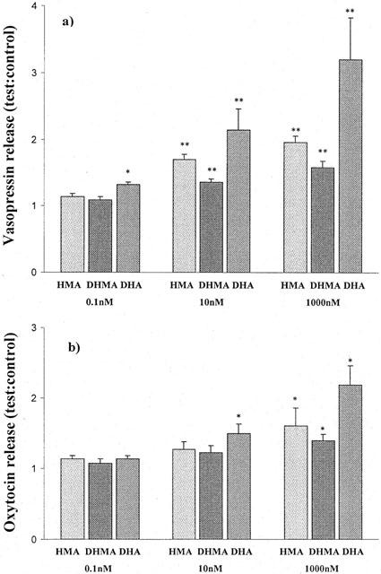
Methylenedioxymethamphetamine (MDMA, 'ecstasy'), widely used as a recreational drug, can produce hyponatraemia. The possibility that this could result from stimulation of vasopressin by MDMA or one of its metabolites has been investigated in vitro. Release of both oxytocin and vasopressin from isolated hypothalami obtained from male Wistar rats was determined under basal conditions and following potassium (40 mM) stimulation. The results were compared with those obtained for basal and stimulated release in the presence of MDMA or metabolites in the dose range 1 microM to 100 pM (n=5 - 8) using Student's t-test with Dunnett's correction for multiple comparisons. All compounds tested affected neurohypophysial hormone release, HMMA (4-hydroxy-3-methoxymethamphetamine) and DHA (3,4-dihydroxyamphetamine) being more active than MDMA, and DHMA (3,4-dihydroxymethamphetamine) being the least active. The effect on vasopressin release was greater than that on oxytocin. In the presence of HMMA the ratio test:control for basal release increased for vasopressin from 1.1+/-0.16 to 2.7+/-0.44 (s.e.m., P<0.05) at 10 nM and for oxytocin from 1.0+/-0.05 to 1.6+/-0.12 in the same hypothalami. For MDMA the ratio increased to 1.5+/-0.27 for vasopressin and to 1.28+/-0.04 for oxytocin for 10 nM. MDMA and its metabolites can stimulate both oxytocin and vasopressin release in vitro, the response being dose dependent for each drug with HMMA being the most potent.
Figures


References
-
- AJAELO I., KOENIG K., SNOEY E. Severe hyponatremia and inappropriate antidiuretic hormone secretion following ecstasy use. Acad. Emerg. Med. 1998;5:839–840. - PubMed
-
- AYUS J.C., WHEELER J.M., ARIEFF A.I. Postoperative hyponatremic encephalopathy in menstruant women. Ann. Intern. Med. 1992;117:891–897. - PubMed
-
- BORGMAN R.J. α-Methyldopamine derivatives. Synthesis and pharmacology. J. Med. Chem. 1974;17:427–430. - PubMed
-
- BECKETT A.H., KIRK G., SHARPEN A.J. The configuration of α-methyldopamine. Tetrahedron. 1965;21:1489–1493.
Publication types
MeSH terms
Substances
LinkOut - more resources
Full Text Sources
Other Literature Sources
Medical
