

Detection of functional connectivity using temporal correlations in MR images
- PMID: 11835612
- PMCID: PMC6872035
- DOI: 10.1002/hbm.10022
Detection of functional connectivity using temporal correlations in MR images
Abstract
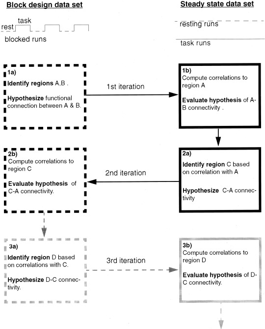
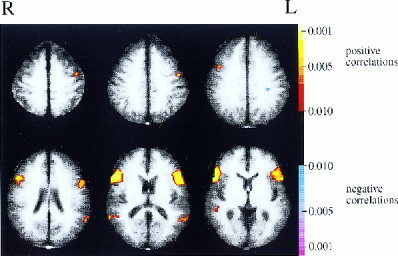
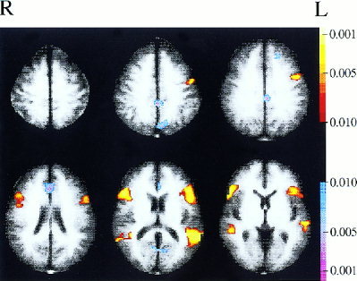
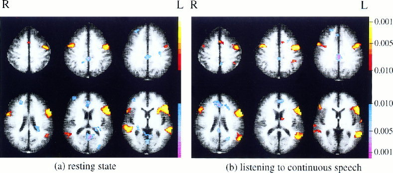
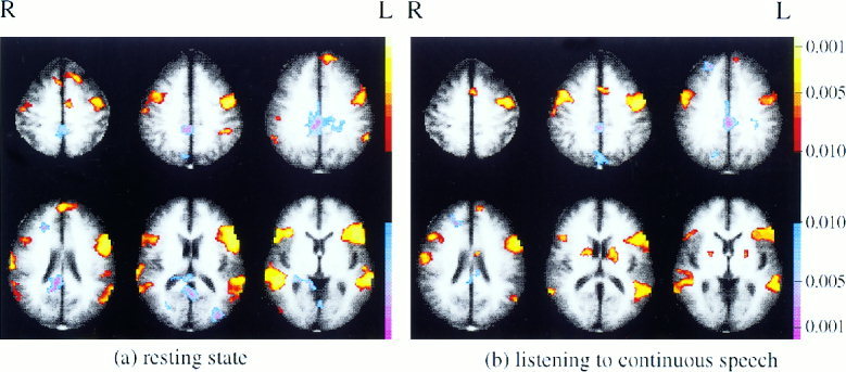
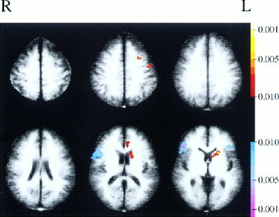
Functional connectivity among brain regions has been investigated via an analysis of correlations between regional signal fluctuations recorded in magnetic resonance (MR) images obtained in a steady state. In comparison with studies of functional connectivity that utilize task manipulations, the analysis of correlations in steady state data is less susceptible to confounds arising when functionally unrelated brain regions respond in similar ways to changes in task. A new approach to identifying interregional correlations in steady state data makes use of two independent data sets. Regions of interest (ROIs) are defined and hypotheses regarding their connectivity are generated in one data set. The connectivity hypotheses are then evaluated in the remaining (independent) data set by analyzing low frequency temporal correlations between regions. The roles of the two data sets are then reversed and the process repeated, perhaps multiple times. This method was illustrated by application to the language system. The existence of a functional connection between Broca's area and Wernicke's area was confirmed in healthy subjects at rest. An increase in this functional connection when the language system was actively engaged (when subjects were continuously listening to narrative text) was also confirmed. In a second iteration of analyses, a correlation between Broca's area and a region in left premotor cortex was found to be significant at rest and to increase during continuous listening. These findings suggest that the proposed methodology can reveal the presence and strength of functional connections in high-level cognitive systems.
Copyright 2002 Wiley-Liss, Inc.
Figures
References
-
- Biswal BB, Hudetz AG, Yetkin FZ, Haughton VM, Hyde JS (1997a): Hypercapnia reversibly suppresses low‐frequency fluctuations in the human motor cortex during rest using echo‐planar MRI. J Cereb Blood Flow Metab 17: 301–308. - PubMed
-
- Biswal BB, Van Kylen J, Hyde JS (1997b): Simultaneous assessment of flow and BOLD signals in resting‐state functional connectivity maps. NMR Biomed 10: 165–170. - PubMed
-
- Biswal BB, Yetkin FZ, Haughton VM, Hyde JS (1995): Functional connectivity in the motor cortex of resting human brain using echo‐planar MRI. Magn Reson Med 34: 537–541. - PubMed
-
- Boatman D, Freeman J, Vining E, Pulsifer M, Miglioretti D, Minahan R, Carson B, Brandt J, McKhann G (1999): Language recovery after left hemispherectomy in children with late‐onset seizures. Ann Neurol 46: 579–586. - PubMed
-
- Boatman D, Hart JJ, Lesser RP, Honeycutt N, Anderson NB, Miglioretti D, Gordon B (1998): Right hemisphere speech perception revealed by amobarbital injection and electrical interference. Neurology 51: 458–464. - PubMed
Publication types
MeSH terms
Grants and funding
LinkOut - more resources
Full Text Sources
Other Literature Sources
Medical
