. 2002 Mar;76(5):2075-86.
doi: 10.1128/jvi.76.5.2075-2086.2002.

A replication-competent, neutralization-sensitive variant of simian immunodeficiency virus lacking 100 amino acids of envelope

Affiliations

A replication-competent, neutralization-sensitive variant of simian immunodeficiency virus lacking 100 amino acids of envelope

Welkin E Johnson et al. J Virol. 2002 Mar.

Abstract

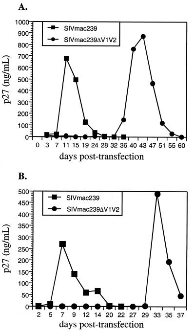
Coding sequences for the first two variable loops of the gp120 envelope glycoprotein were removed from simian immunodeficiency virus (SIV) strain 239 (SIVmac239). This deletion encompassed 100 amino acids. The resulting virus replicated poorly after transfection into immortalized T-cell lines, with peak replication occurring only after 25 to 30 days. Limited passaging of SIVmac239DeltaV1V2 in cultures gave rise to a variant which had significantly improved replication kinetics but which retained the original 100-amino-acid deletion in gp120. Cloning and sequencing revealed 11 changes in the envelope, including amino acid substitutions in both gp120 (5 substitutions) and gp41(6 substitutions). Four of the five changes in gp120 are predicted to lie within and around the putative coreceptor binding domain, a region which is believed to be covered by the V1 and V2 loops in the native envelope complex. Analysis of recombinant clones surprisingly revealed that the changes in gp41 were sufficient to overcome the replication deficiency created by deletion of the V1 and V2 loops from gp120. The SIVmac239DeltaV1V2 envelope displayed a significant reduction in its ability to mediate cell-cell fusion, and the infectious titer of SIVmac239DeltaV1V2 was approximately four- to eightfold lower than that of parental SIVmac239. Although SIVmac239 is strongly dependent on both CD4 and a coreceptor for entry, envelope protein lacking the V1 and V2 loops was able to mediate fusion with CD4(-) CCR5(+) cells at 60% the level observed with CD4(+) CCR5(+) cells. Plasma from SIVmac239-infected monkeys was at least 100 to 1,000 times more effective at neutralizing SIVmac239DeltaV1V2 than SIVmac239. These results demonstrate the dispensability of the V1-V2 sequences of SIVmac239 for viral replication, a role for V1 and V2 in shielding the coreceptor binding region of the envelope, and the extreme sensitivity of a SIV lacking these sequences to antibody-mediated neutralization.


Figures

FIG. 1.
FIG. 1.
Variable loops V1 and V2. (A) Folding pattern of the V1 and V2 loops of HIV-1 Env protein gp120. (B) Predicted folding pattern of the V1-V2 structure of SIVmac239 (12). (C) SIVmac239ΔV1V2. The deletion mutant was created by deleting 100 amino acids (a.a.) of envelope, encompassing all of V1 and V2, and replacing the deleted residues with a Gly-Ala-Gly linker. The deletion mutant retains the stem of the V1-V2 structure. Potential sites of N-linked glycosylation attachment are indicated by asterisks.
FIG. 2.
FIG. 2.
Replication of SIVmac239ΔV1V2. Mutant DNA was used to transfect immortalized rhesus macaque T cells (221 cells), and viral replication was assayed by monitoring the expression of p27 capsid protein in the supernatant. Panels A and B show results of two independent transfections.
FIG. 3.
FIG. 3.
Infectious titer of SIVmac239ΔV1V2. Viral stocks were normalized for p27 content and then used for duplicate infections of CEMx174SIV-SEAP indicator cells, which contain a Tat-inducible gene for SEAP. SEAP activity was measured 72 h postinfection, a measure which principally reflects the first round of infection (26). Indicator cells were the same cells as those used in the neutralization assay (see Materials and Methods). Error bars show standard deviations.
FIG. 4.
FIG. 4.
The mutant envelope complex with a deletion of V1 and V2 can mediate CD4-independent fusion. Effector cells expressed parental SIVmac239 envelope (black bars), SIVmac316 envelope (grey bars), or SIVmac239ΔV1V2 envelope (white bars). Target cells expressed CD4 and coreceptor CCR5, CD4 alone, or CCR5 alone. Error bars show standard deviations. (For more details, see Materials and Methods.)
FIG. 5.
FIG. 5.
Infectivity of passaged SIVmac239ΔV1V2. SIVmac239ΔV1V2 was passaged continuously on 221 cells, and supernatants were collected weekly. Supernatants collected on days 85 (d85), 119, and 133 of passage were normalized for p27 content and used to infect fresh 221 cells.
FIG. 6.
FIG. 6.
Mapping of compensatory changes in SIVmac239ΔV1V2(d119). (A) The top two lines depict the parental and mutant constructs; arrows indicate restriction fragments used to generate subclones. The third line depicts the envelope gene cloned from day 119 posttransfection, with the positions of the 11 nonsynonymous changes indicated. The last six lines depict the subclones used to map changes that compensate for the deletion of the V1 and V2 loops. The relative capacity of each variant to replicate in 221 cells is indicated to the right (++++, replication like that of the wild type; −, no detectable replication; −/+, extreme delay in replication typical of SIVΔV1V2 transfections; according to the data in Fig. 6B and other results not shown). (B) Replication of subclones of SIVmac239ΔV1V2(d119) in 221 cells. Data for variant X-C/M-N are not shown.
FIG. 6.
FIG. 6.
Mapping of compensatory changes in SIVmac239ΔV1V2(d119). (A) The top two lines depict the parental and mutant constructs; arrows indicate restriction fragments used to generate subclones. The third line depicts the envelope gene cloned from day 119 posttransfection, with the positions of the 11 nonsynonymous changes indicated. The last six lines depict the subclones used to map changes that compensate for the deletion of the V1 and V2 loops. The relative capacity of each variant to replicate in 221 cells is indicated to the right (++++, replication like that of the wild type; −, no detectable replication; −/+, extreme delay in replication typical of SIVΔV1V2 transfections; according to the data in Fig. 6B and other results not shown). (B) Replication of subclones of SIVmac239ΔV1V2(d119) in 221 cells. Data for variant X-C/M-N are not shown.
FIG. 7.
FIG. 7.
(A) Surface expression of the envelope. 293T cells were transfected with Env expression constructs, and cells were harvested 48 h posttransfection, stained with monoclonal antibody KK42 and a fluorescein isothiocyanate-conjugated secondary antibody, and then sorted by flow cytometry. (B) Cell-cell fusion assay. 293T cells were transfected with envelope expression constructs as in panel A or control vectors. At 48 h posttransfection, the cells were overlaid with CEMx174SIV-SEAP indicator cells, and SEAP activity was measured at the indicated times. (See Materials and Methods for details.)
FIG. 8.
FIG. 8.
Neutralization of SIVmac239ΔV1V2 by SIV-positive rhesus monkey plasma. Increasing concentrations of stock SIVmac239 or SIVmac239ΔV1V2 were incubated with control plasma (squares) or with pooled SIV-positive plasma (circles) diluted 1:200 with RPMI medium. (A) Effect of increasing concentrations of SIVmac239 on sensitivity to SIV-positive plasma. (B) Effect of increasing concentrations of SIVmac239ΔV1V2 on sensitivity to SIV-positive plasma. (C) Effect of increasing concentrations of SIVmac239ΔV1V2(clone C-N) on sensitivity to SIV-positive plasma.
FIG. 9.
FIG. 9.
SIVmac239ΔV1V2 is extremely sensitive to neutralization by SIV-positive plasma. CEMx174SIV-SEAP cells were infected with virus in the presence of increasing dilutions of plasma from uninfected rhesus macaques (closed symbols) or plasma from SIV-positive rhesus macaques (open symbols). (A) Neutralization of SIVmac239ΔV1V2. Background SEAP levels in this experiment are indicated by the line labeled “uninfected.” (B) Comparative neutralization of SIVmac239ΔV1V2 and SIVmac239ΔV1V2(clone C-N).
FIG. 10.
FIG. 10.
Neutralization of SIVmac239ΔV1V2 with various plasma samples. Plasma samples from three SIV-positive rhesus monkeys were tested for neutralizing activity against SIVmac239 (open symbols) and SIVmac239ΔV1V2 (closed symbols). (A) Plasma collected from animals at week 16 postinfection. (B) Plasma collected at week 24 postinfection.

References

    1. Alexander, W. A., B. Moss, and T. R. Fuerst. 1992. Regulated expression of foreign genes in vaccinia virus under the control of bacteriophage T7 RNA polymerase and the Escherichia coli lac repressor. J. Virol. 66:2934-2942. - PMC - PubMed
    1. Ausubel, F. M., R. Brent, R. E. Kingston, D. D. Moore, J. G. Seidman, J. A. Smith, and K. Struhl (ed.). 1987. Current protocols in molecular biology. John Wiley & Sons, Inc., New York, N.Y.
    1. Bandres, J. C., Q. F. Wang, J. O'Leary, F. Baleaux, A. Amara, J. A. Hoxie, S. Zolla-Pazner, and M. K. Gorny. 1998. Human immunodeficiency virus (HIV) envelope binds to CXCR4 independently of CD4, and binding can be enhanced by interaction with soluble CD4 or by HIV envelope deglycosylation. J. Virol. 72:2500-2504. - PMC - PubMed
    1. Bosch, M. L., P. L. Earl, K. Fargnoli, S. Picciafuoco, F. Giombini, F. Wong-Staal, and G. Franchini. 1989. Identification of the fusion peptide of primate immunodeficiency viruses. Science 244:694-697. - PubMed
    1. Burns, D. P., C. Collignon, and R. C. Desrosiers. 1993. Simian immunodeficiency virus mutants resistant to serum neutralization arise during persistent infection of rhesus monkeys. J. Virol. 67:4104-4113. - PMC - PubMed

Publication types

MeSH terms