. 2002 Mar;76(5):2159-67.
doi: 10.1128/jvi.76.5.2159-2167.2002.

Degradation of STAT1 and STAT2 by the V proteins of simian virus 5 and human parainfluenza virus type 2, respectively: consequences for virus replication in the presence of alpha/beta and gamma interferons

Affiliations

Degradation of STAT1 and STAT2 by the V proteins of simian virus 5 and human parainfluenza virus type 2, respectively: consequences for virus replication in the presence of alpha/beta and gamma interferons

J Andrejeva et al. J Virol. 2002 Mar.

Abstract

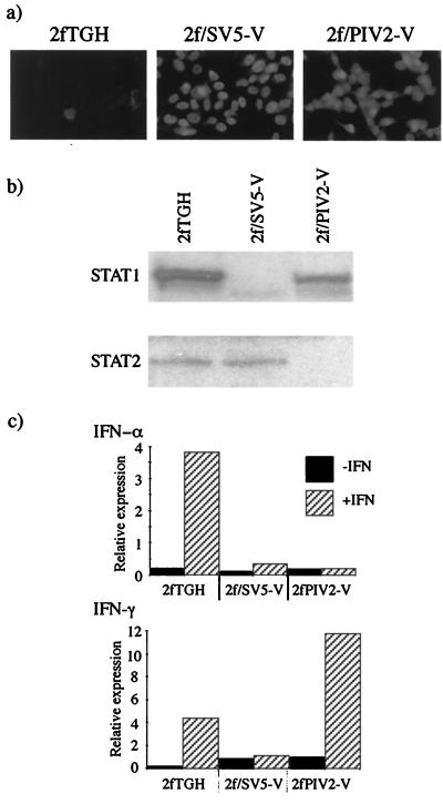
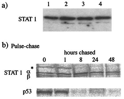
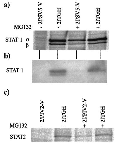
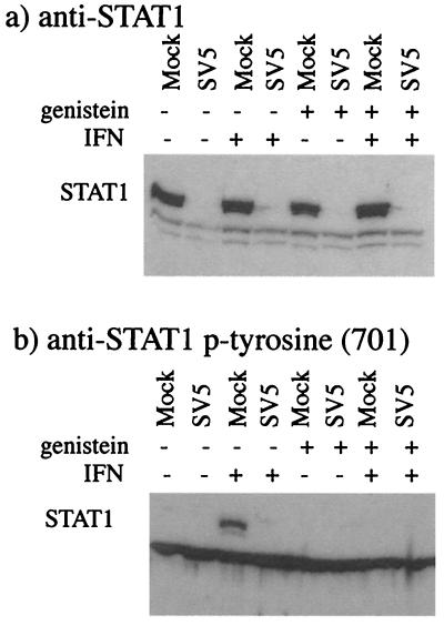
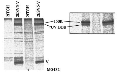
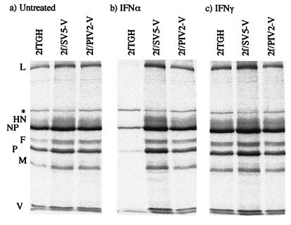
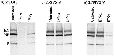
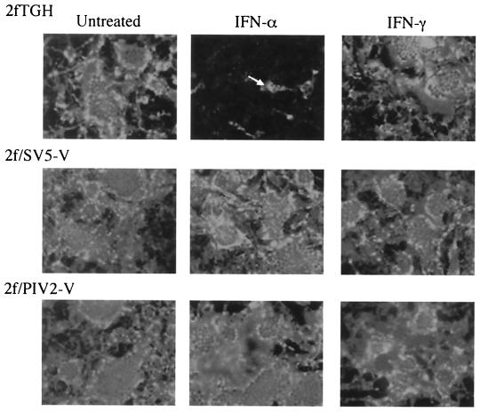
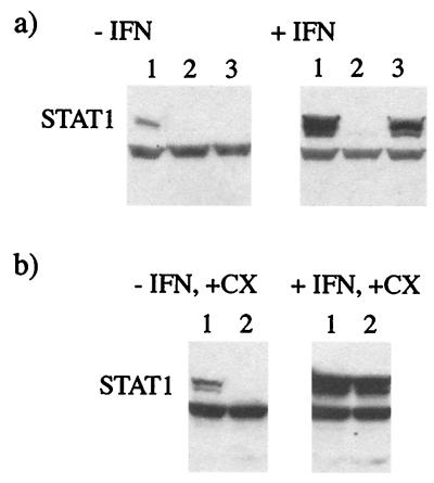
Human cell lines were isolated that express the V protein of either simian virus 5 (SV5) or human parainfluenza virus type 2 (hPIV2); the cell lines were termed 2f/SV5-V and 2f/PIV2-V, respectively. STAT1 was not detectable in 2f/SV5-V cells, and the cells failed to signal in response to either alpha/beta interferons (IFN-alpha and IFN-beta, or IFN-alpha/beta) or gamma interferon (IFN-gamma). In contrast, STAT2 was absent from 2f/PIV2-V cells, and IFN-alpha/beta but not IFN-gamma signaling was blocked in these cells. Treatment of both 2f/SV5-V and 2f/PIV2-V cells with a proteasome inhibitor allowed the respective STAT levels to accumulate at rates similar to those seen in 2fTGH cells, indicating that the V proteins target the STATs for proteasomal degradation. Infection with SV5 can lead to a complete loss of both phosphorylated and nonphosphorylated forms of STAT1 by 6 h postinfection. Since the turnover of STAT1 in uninfected cells is longer than 24 h, we conclude that degradation of STAT1 is the main mechanism by which SV5 blocks interferon (IFN) signaling. Pretreatment of 2fTGH cells with IFN-alpha severely inhibited both SV5 and hPIV2 protein synthesis. However, and in marked contrast, pretreatment of 2fTGH cells with IFN-gamma had little obvious effect on SV5 protein synthesis but did significantly reduce the replication of hPIV2. Pretreament with IFN-alpha or IFN-gamma did not induce an antiviral state in 2f/SV5-V cells, indicating either that the induction of an antiviral state is completely dependent on STAT signaling or that the V protein interferes with other, STAT-independent cell signaling pathways that may be induced by IFNs. Even though SV5 blocked IFN signaling, the addition of exogenous IFN-alpha to the culture medium of 2fTGH cells 12 h after a low-multiplicity infection with SV5 significantly reduced the subsequent cell-to-cell spread of virus. The significance of the results in terms of the strategy that these viruses have evolved to circumvent the IFN response is discussed.


Figures

FIG. 1.
FIG. 1.
Characterization of 2f/SV5-V and 2f/PIV2-V cells. (a) Photomicrographs showing the intracellular localization of the V proteins of SV5 and hPIV2 in 2f/SV5-V and 2f/PIV2-V cells, respectively. Note that all the cells are positive for V and that V has a primarily nuclear distribution. (b) Immunoblot analysis demonstrating the presence of STAT2 but the absence of STAT1 in 2f/SV5-V cells and the presence of STAT1 but the absence of STAT2 in 2f/PIV2-V cells. Total extracts of 2f/SV5-V and 2f/PIV2-V cells were electrophoresed through 10% polyacrylamide gels and electrophoretically transferred to nitrocellulose, and STAT1 and STAT2 were detected by immunoblot analysis. (c) IFN-α signaling is inhibited in 2f/SV5-V and 2f/PIV2-V cells (top panel), while IFN-γ signaling is inhibited in 2f/SV5-V but not 2f/PIV2-V cells (bottom panel). Cells were transfected with either IFN-α- or IFN-γ-responsive plasmids together with pJATlacZ, and at 46 h posttransfection, cells were either treated with human IFN-α or IFN-γ or left untreated. Four hours later, luciferase and β-galactosidase activities in the cellular lysates were measured. Luciferase activity, expressed in relative light units, was normalized to β-galactosidase activity.
FIG. 2.
FIG. 2.
STAT1 has a slow intracellular turnover rate. (a) STAT1 was detected by immunoblot analysis in total extracts of 2fTGH cells that had been treated with cycloheximide (50 μg/ml) for 0, 4, 8, or 18 h (lanes 1 to 4, respectively). (b) 2fTGH cells were radioactively labeled with 35[S]methionine for 1 h and then cultured (chased) for 0, 1, 8, 24, or 48 h in medium that did not contain 35[S]methionine. Soluble antigen extracts were made, and STAT1α and -β and p53 were immunoprecipitated. An asterisk indicates an unidentified host cell protein that is rapidly turned over.
FIG. 3.
FIG. 3.
The proteasome inhibitor MG132 blocks the degradation of newly synthesized STAT1 in 2f/SV5-V cells and STAT2 in 2f/PIV2-V cells. (a and c) MG132 (10 μM) was or was not added to the culture 1 h before the cells were radioactively labeled with 35[S]methionine for a further 7 h in the presence or absence of the inhibitor. STAT1α and -β were immunoprecipitated from 2f/SV5-V soluble antigen extracts (panel a), and STAT2 was immunoprecipitated from 2f/PIV2-V extracts (panel c). (b) Immunoblot analysis was also used to detect STAT1 in samples of total extracts of the 2f/SV5-V cells used in the immunoprecipitation analysis.
FIG. 4.
FIG. 4.
Both phosphorylated and nonphoshorylated forms of STAT1 are degraded by SV5. 2fTGH cells were or were not pretreated with the kinase inhibitor genistein (100 μM) for 1 h prior to being mock infected or infected with SV5 at an MOI of 10. Cells were or were not cultured in the presence of genistein for a further 6 h. Total cell extracts were made, and the presence of STAT1 was detected by immunoblot analysis with polyclonal anti-STAT1 antibody (a) or anti- phosphotyrosine (Y701) STAT1 [anti-STAT1 p-tyrosine (701)] (b).
FIG. 5.
FIG. 5.
Coimmunoprecipitation of host cell proteins with the V protein of SV5. 2fTGH and 2f/SV5-V cells were or were not treated with the proteasome inhibitor MG132 (10 μM) for 6 h, during which time they were also metabolically labeled with 35[S]methionine. Soluble antigen extracts were made, and the V protein was precipitated with MAb SV5-P-k. The precipitated polypeptides were separated on both 12 and 7% (inset) polyacrylamide gels. The lower of the two high-molecular-mass host cell bands that coprecipitated with V was identified by MALDI-TOF analysis as the 127-kDa subunit of UV DDB; the upper, 150-kDa band has yet to be identified (unpublished results).
FIG. 6.
FIG. 6.
Effect of IFN pretreatment on SV5 protein synthesis in 2fTGH, 2f/SV5-V, and 2f/PIV2-V cells. Cells were not (a) or were pretreated with either IFN-α (b) or IFN-γ (c) (1,000 IU/ml) for 16 h prior to infection with SV5 at an MOI of 0.5 to 1.0 PFU/cell. At 20 h p.i., the cells were metabolically labeled with 35[S]methionine for 1 h. Virus proteins were precipitated from soluble antigen extracts of these cells with a pool of MAbs to the NP, P, M, HN, and F proteins; separated by 10% PAGE; and visualized by phosphorimager analysis. An asterisk indicates an unidentified host cell protein.
FIG. 7.
FIG. 7.
Effect of IFN pretreatment on hPIV2 protein synthesis in 2fTGH (a), 2f/SV5-V (b), and 2f/PIV2-V (c) cells. Cells were or were not pretreated with either IFN-α or IFN-γ (1,000 IU/ml) for 16 h prior to infection with hPIV2 at an MOI of 0.5 to 1.0 PFU/cell. At 20 h p.i., the cells were metabolically labeled with 35[S]methionine for 1 h. Virus proteins were precipitated from soluble antigen extracts of these cells with a pool of MAbs to the NP, P, and HN proteins; separated by 10% PAGE; and visualized by phosphorimager analysis.
FIG. 8.
FIG. 8.
Photomicrographs demonstrating the cell-to-cell spread of SV5 in 2fTGH, 2f/SV5-V, and 2f/PIV2-V cells in the presence or absence of exogenous IFN-α or IFN-γ. Cells were infected at 0.01 PFU/cell; 12 h later, IFN-α or IFN-γ (1,000 IU/ml) was or was not added to the culture medium. Monolayers were fixed at various times p.i., and the percentage of infected cells was estimated by immunofluorescence with a pool of MAbs to the NP and P proteins. At 24 h p.i., >5% of the cells in all the monolayers were infected (data not shown). By 72 h p.i., all the cells were infected, except for 2fTGH cells that had been cultured in the presence of IFN-α. For reference, a single cell is highlighted with a white arrow.
FIG. 9.
FIG. 9.
De novo SV5 protein synthesis is required for STAT1 degradation in IFN-pretreated cells but not in untreated cells. (a) 2fTGH cells were (+IFN) or were not (−IFN) pretreated with IFN-α (1,000 IU ml) for 18 h prior to being mock infected (lane 1) or infected with “infectious” SV5 (lane 2) or UV-inactivated virus (lane 3; the efficiency of UV inactivation was demonstrated by showing that even after 24 h of infection, no virus protein synthesis had occurred, as judged by immunofluorescence). At 8 h p.i., total cell extracts were made, and STAT1 was detected by immunoblot analysis. (b) 2fTGH cells were (+IFN) or were not (−IFN) pretreated with IFN-α for 18 h prior to being mock infected (lane 1) or infected with “infectious” SV5 (lane 2). From the time of infection, cells were cultured in the presence of cycloheximide (+CX; 50 μg/ml). At 8 h p.i., total cell extracts were made, and STAT1 was detected by immunoblot analysis.

References

    1. Choppin, P. W. 1964. Multiplication of a myxovirus (SV5) with minimal cytopathic effects and without interference. Virology 23:224-233. - PubMed
    1. Didcock, L., D. F. Young, S. Goodbourn, and R. E. Randall. 1999. Sendai virus and simian virus 5 block activation of interferon-responsive genes: importance for virus pathogenesis. J. Virol. 73:3125-3133. - PMC - PubMed
    1. Didcock, L., D. F. Young, S. Goodbourn, and R. E. Randall. 1999. The V protein of simian virus 5 inhibits interferon signaling by targeting STAT1 for proteasome-mediated degradation. J. Virol. 73:9928-9933. - PMC - PubMed
    1. Garcin, D., P. Latorre, and D. Kolakofsky. 1999. Sendai virus C proteins counteract the interferon-mediated induction of an antiviral state. J. Virol. 73:6559-6565. - PMC - PubMed
    1. Garcin, D., J. Curran, and D. Kolakofsky. 2000. Sendai virus C proteins must interact directly with cellular components to interfere with interferon action. J. Virol. 74:8823-8830. - PMC - PubMed

Publication types

MeSH terms