. 2002 Mar;76(5):2263-73.
doi: 10.1128/jvi.76.5.2263-2273.2002.

Human papillomavirus type 31 replication modes during the early phases of the viral life cycle depend on transcriptional and posttranscriptional regulation of E1 and E2 expression

Affiliations

Human papillomavirus type 31 replication modes during the early phases of the viral life cycle depend on transcriptional and posttranscriptional regulation of E1 and E2 expression

Walter G Hubert et al. J Virol. 2002 Mar.

Abstract

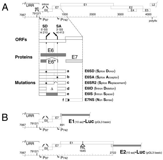
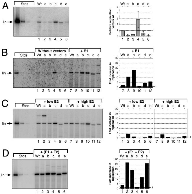
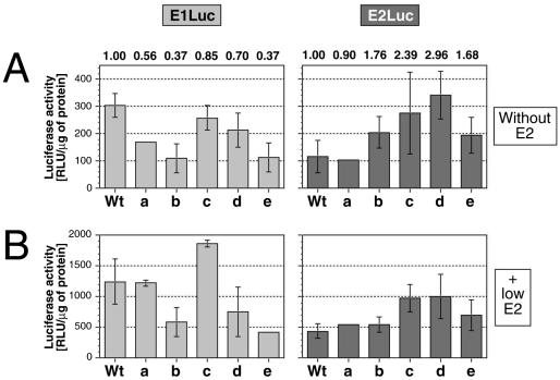
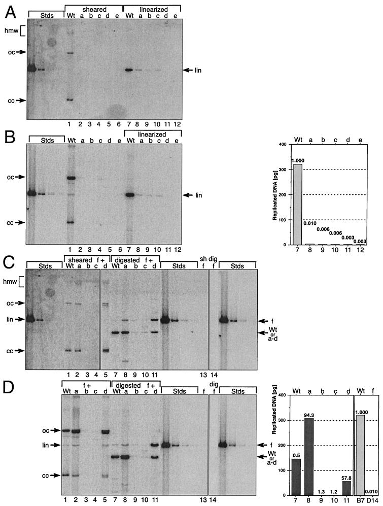
The E1 and E2 proteins are both required for papillomavirus DNA replication, and replication efficiency is controlled by the abundance of these factors. In human papillomaviruses (HPVs), the regulation of E1 and E2 expression and its effect on viral replication are not well understood. In particular, it is not known if E1 and E2 modulate their own expression and how posttranscriptional mechanisms may affect the levels of the replication proteins. Previous studies have implicated splicing within the E6 open reading frame (ORF) as being important for modulating replication of HPV type 31 (HPV31) through altered expression of E1 and E2. To analyze the function of the E6 intron in viral replication more specifically, we examined the effects of E6 splicing mutations in the context of entire viral genomes in transient assays. HPV31 genomes which had mutations in the splice donor site (E6SD) or the splice acceptor site (E6SA), a deletion of the intron (E6ID), or substituted heterologous intron sequences (E6IS) were constructed. Compared to wild-type (wt) HPV31, pHPV31-E6SD, -E6SA, and -E6IS replicated inefficiently while pHPV31-E6ID replicated at an intermediate level. Cotransfection of the E6 mutant genomes with an E1 expression vector strongly activated their replication levels, indicating that efficient expression of E1 requires E6 internal splicing. In contrast, replication was activated only moderately with an E2 expression vector. Replacing the wt E6 intron in HPV31 with a heterologous intron from simian virus 40 (E6SR2) resulted in replication levels similar to that of the wt in the absence of expression vectors, suggesting that mRNA splicing upstream of the E1 ORF is important for high-level replication. To examine the effects of E6 intron splicing on E1 and E2 expression directly, we constructed reporter DNAs in which the luciferase coding sequences were fused in frame to the E1 (E1Luc) or E2 (E2Luc) gene. Reporter activities were then analyzed in transient assays with cotransfected E1 or E2 expression vectors. Both reporters were moderately activated by E1 in a dose-dependent manner. In addition, E1Luc was activated by low doses of E2 but was repressed at high doses. In contrast, E2 had little effect on E2Luc activity. These data indicate that E1 expression and that of E2 are interdependent and regulated differentially. When the E6 splicing mutations were analyzed in both reporter backgrounds, only E1Luc activities correlated with splicing competence in the E6 ORF. These findings support the hypothesis that the E6 intron primarily regulates expression of E1. Finally, in long-term replication assays, none of the E6 mutant genomes could be stably maintained. However, cotransfection of the E6 splicing mutant genomes with pHPV31-E7NS, which contains a nonsense mutation in the E7 coding sequence, restored stable replication of some mutants. Our observations indicate that E1 expression and that of E2 are differentially regulated at multiple levels and that efficient expression of E1 is required for transient and stable viral replication. These regulatory mechanisms likely act to control HPV copy number during the various phases of the viral life cycle.


Figures

FIG. 1.
FIG. 1.
Diagrams for plasmids and mutations used in this study. (A) Early region of the HPV31 genome (top) with the URR, replication origin (ori), ORFs (open boxes), promoters (horizontal arrows), and selected mRNA splice sites (slanted arrows). The enlargement (bottom) shows ORFs E6 and E7 (open boxes), their proteins (stippled boxes), and the location of the mutations. Nucleotide positions refer to HPV31 unless stated otherwise. a, inactivated splice donor (nt 212T→C); b, inactivated splice acceptor (nt 411A→C); c, inserted splice replacement (HPV31 nt 344→415 replaced with SV40 small t antigen intron from pGL2 control [Promega; pGL2 nt 2157→2228], nt 212T→C, and nt 342T→C); d, E6 intron deletion including nt 211→412; e, inserted E6 intron spacer replacing HPV31 nt 344→415 with amino phosphotransferase coding sequences from pREP10 (Stratagene; pREP10, nt 248→449); f, E7 nonsense mutation (nt 610T→C and nt 611G→T, E7 Glu18→amber). (B) Schematic maps of HPV31 E1- and E2-specific luciferase reporters E1Luc and E2Luc, respectively, in the pGL3 basic parental plasmid (Promega). In each reporter, the firefly luciferase gene is fused in frame to the first 10 codons of the specified HPV gene. E1Luc contains nt 7067 to 892 from HPV31. E2Luc contains nt 7067 to 2723 from HPV31. The E1 gene in E2Luc is inactivated with a six-frame translational termination linker at nt 1645. aa, amino acids.
FIG. 2.
FIG. 2.
The effects of splicing mutations in the E6 ORF on transient replication of HPV31 genomes. SCC13 cells were either transfected with ligated viral DNAs by themselves or cotransfected with HPV31 DNA and constant amounts of expression vectors for E1 or E2 or both. The amounts of replicated DNAs were quantified and graphed for each panel. Wt, wt HPV31. Mutations in the HPV31 genome background: a, pHPV31-E6SD; b, pHPV31-E6SA; c, pHPV31-E6SR2; d, pHPV31-E6ID; e, pHPV31-E6IS. Each autoradiogram contains HPV31 standards (Stds): 500, 25, 2.5, and 0.5 pg of linearized DNA (7,912 bp) (lin, migratory position in 0.8% agarose gels). The DNA bands migrating more slowly than the linearized samples in panels B through D are due to multimeric forms generated during the initial ligation and transfection. (A) Autoradiogram of replicating of viral genomes without expression vectors. Graphs indicate the relative replication levels of mutants versus wt and represent the averages ± standard deviations of samples quantified from panels A and B, lanes 1 through 6. (B) Autoradiogram of replicating viral genomes without expression vectors (lanes 1 through 6) or in the presence of equimolar amounts (versus the HPV genome) of the E1 expression vector (lanes 7 through 12). Bars 7 through 12 indicate how the replication level of each DNA is increased by the cotransfected E1 vector. (C) Autoradiogram of replicating viral genomes in the presence of small (0.1 M) or large (equimolar) amounts of the E2 expression vector. The graph indicates how the replication level of each DNA is increased by the small (bars 1 through 6) and large (bars 7 through 12) amounts of cotransfected E2 vector. (D) Autoradiogram of replicating viral genomes with equimolar amounts of E1 and E2 expression vectors. The graph indicates how the replication level of each DNA is increased by the presence of both vectors.
FIG. 3.
FIG. 3.
The response of E1Luc and E2Luc reporters to increasing amounts of E1 or E2. Shown are activities from transient reporter assays. SCC13 cells were cotransfected with constant amounts of reporter DNA and increasing amounts of either an E1 or E2 expression vector. Mutations in both reporter backgrounds: a, E6SD; b, E6SA; c, E6SR2; d, E6ID; e, E6IS. Each datum represents the average specific luciferase activity of two independent transfections (relative light units[RLU] per microgram of lysate protein) versus the molar ratio of expression vector to reporter DNA. Error bars, standard deviations (at some data points, bars are obscured by the symbol). The value labels show the relative activities versus that for transfection without the expression vector (basal activity). To facilitate graphing on a log scale, the datum of the zero expression vector was assigned a molar ratio of 0.001. (A) Dose response of E1Luc versus E1 (0 to 10 molar ratio). (B) Dose response of E1Luc versus E2 (0 to 5 molar ratio). (C) Dose response of E2Luc versus E1 (0 to 10 molar ratio). (D) Dose response of E2Luc versus E2 (0 to 5 molar ratio).
FIG. 4.
FIG. 4.
Response of E1Luc and E2Luc reporters to the combination of E1 and E2. SCC13 cells were transfected in transient assays, and the resulting reporter activities were graphed. All cotransfection mixtures contained a constant amount of reporter DNA (E1Luc or E2Luc) and increasing amounts of one expression vector (E1 or E2 vector). In addition, transfections were performed either without or with a constant amount of the second expression vector as indicated (molar ratio versus reporter). Mutations in both reporter backgrounds: a, E6SD; b, E6SA; c, E6SR2; d, E6ID; e, E6IS. In each panel, the graph shows the specific luciferase activities resulting from transfections with three DNAs (reporter plus E1 and E2 DNAs [solid lines and symbols]) or with two DNAs (reporter plus one vector [dashed lines and open symbols]) to assess the contribution of replication to reporter activity. Each datum represents the average specific luciferase activity of two independent transfections (relative light units [RLU] per microgram of lysate protein) versus the molar ratio of expression vector versus reporter DNA. Error bars, standard deviations (at some data points, bars are obscured by symbols). To facilitate graphing on a log scale, the datum of the zero expression vector was assigned a molar ratio of 0.001. (A) Dose response of E1Luc versus E1 (0 to 10 molar ratio) in the absence or presence of constant 0.1 M amounts of E2 vector. (B) Dose response of E1Luc versus E2 (0 to 5 molar ratio) in the absence or presence of constant 0.2 M amounts of E1 vector. (C) Dose response of E2Luc versus E1 (0 to 10 molar ratio) in the absence or presence of constant 0.1 M amounts of E2 vector. (D) Dose response of E2Luc versus E2 (0 to 5 molar ratio) in the absence or presence of constant 0.2 M amounts of E1 vector.
FIG. 5.
FIG. 5.
The response of E1Luc and E2Luc reporters containing mutations in the E6 splice sites to increasing amounts of E1 or E2. The graphs show the specific E1Luc and E2Luc reporter activities from transient transfection assays with SCC13 cells. Transfection mixtures contained a reporter DNA, and transfections were performed without the E2 vector (basal) or with a 0.1 molar ratio of E2 expression vector as indicated. Mutations in both reporter backgrounds: a, E6SD; b, E6SA; c, E6SR2; d, E6ID; e, E6IS. Each datum represents the average specific luciferase activity of two independent transfections (relative light units [RLU] per microgram of lysate protein). Error bars, standard deviations. (A) Absolute basal activities of the E1Luc and E2Luc reporters. The value labels show the relative activities of mutants versus wt in each reporter background. (B) Absolute E2-induced activities of the E1Luc and E2Luc reporters in the presence of 0.1 M amounts of the E2 expression vector.
FIG. 6.
FIG. 6.
Stable replication of HPV31 genomes containing mutant E6 splice sites. Shown is the stable replication of wt and mutant HPV31 genomes: Wt, wt HPV31; a, pHPV31-E6SD; b, pHPV31-E6SA; c, pHPV31-E6SR2; d, pHPV31-E6ID; e, pHPV31-E6IS. Primary HFK were stably transfected with individual viral DNAs (A and B) or paired DNAs (C and D), where the wt and mutants a through e were each cotransfected with mutant f (pHPV31-E7NS). Each autoradiogram contains HPV31 standards (Stds): 500, 25, 2.5, and 0.5 pg of linearized DNA (7,912 bp). Aliquots (5 μg) of total cellular DNA were analyzed in panels A and C; therefore, the DNA standards represent about 100, 5, 0.25, and 0.1 viral genomes per cell, respectively. Arrows, migration of the different topological forms of the viral DNA. A high-molecular-weight (hmw) smear indicates that the HPV DNA is either integrated into the cellular genome or replicates as a plasmid multimer. The covalently closed (cc) and open (oc) formsof HPV DNA result from autonomously replicating plasmid monomers. Linearized (lin) HPV DNA bands were used to quantify the replicated viral DNA. (A) Autoradiogram from transfections with individual HPV31 genomes (wt and mutants a through e). DpnI-resistant, sheared-total-DNA samples are shown on the left (lanes 1 to 6), and the linearized ones are shown on the right (lanes 7 to 12). (B) Autoradiogram showing low-molecular-weight (Hirt) DNA isolated and analyzed from the same cell lines as in panel A. DpnI-resistant samples are shown on the left (lanes 1 to 6), and linearized ones are shown on the right (lanes 7 to 12). The linearized sample bands were quantified and graphed. The value labels indicate the relative replication levels of mutants versus wt. (C) Autoradiogram from transfections with paired HPV31 genomes (wt and mutants a through d, each paired with f). Lanes 1 to 5, DpnI-resistant-, sheared-total-DNA samples; lanes 7 to 12, digested-DNA samples; lanes 13 (sheared) and 14 (digested), stable replication of pHPV31-E7NS (f) by itself. Arrows, positions of pHPV31-E7NS (f) and the cotransfected DNA (wt and a to d). (D) Autoradiogram showing low-molecular-weight (Hirt) DNA isolated and analyzed from the same cell lines as in panel C. DpnI-resistant samples (left; lanes 1 to 5) and digested ones (middle; lanes 7 to 11) are shown. Lanes 13 and 14, stable replication of pHPV31-E7NS (f) by itself (digested). Arrows, positions of pHPV31-E7NS (f) and cotransfected DNA (wt and a to d). The digested lower sample bands (wt, a to d) were quantified and graphed (right). The value labels show the relative increases of stable replication levels of the sample DNAs with pHPV31-E7NS (f) compared to the autonomous levels (panel B). The replication of wt (panel B, lane 7) and pHPV31-E7NS (panel D, lane 14) in single DNA transfections are compared (far right). The value labels in graphs 7 and 14 indicate the relative replication levels of pHPV31-E7NS versus wt.

Similar articles

Cited by

References

    1. Belaguli, N. S., M. M. Pater, and A. Pater. 1995. Splice sites of human papillomavirus type 16 E6 gene or heterologous gene required for transformation by E7 and accumulation of E7 RNA. J. Med. Virol. 47:445-453. - PubMed
    1. Butz, K., and F. Hoppe-Seyler. 1993. Transcriptional control of human papillomavirus (HPV) oncogene expression: composition of the HPV type 18 upstream regulatory region. J. Virol. 67:6476-6486. - PMC - PubMed
    1. Chiang, C. M., G. Dong, T. R. Broker, and L. T. Chow. 1992. Control of human papillomavirus type 11 origin of replication by the E2 family of transcription regulatory proteins. J. Virol. 66:5224-5231. - PMC - PubMed
    1. Chiang, C. M., M. Ustav, A. Stenlund, T. F. Ho, T. R. Broker, and L. T. Chow. 1992. Viral E1 and E2 proteins support replication of homologous and heterologous papillomaviral origins. Proc. Natl. Acad. Sci. USA 89:5799-5803. - PMC - PubMed
    1. Chow, L. T., T. R. Broker, and J. B. Lewis. 1979. Complex splicing patterns of RNAs from the early regions of adenovirus-2. J. Mol. Biol. 134:265-303. - PubMed

Publication types

MeSH terms

Substances

LinkOut - more resources