. 2002 Mar;76(5):2329-39.
doi: 10.1128/jvi.76.5.2329-2339.2002.

The tRNA primer activation signal in the human immunodeficiency virus type 1 genome is important for initiation and processive elongation of reverse transcription

Affiliations

The tRNA primer activation signal in the human immunodeficiency virus type 1 genome is important for initiation and processive elongation of reverse transcription

Nancy Beerens et al. J Virol. 2002 Mar.

Abstract

Human immunodeficiency virus type 1 (HIV-1) reverse transcription is primed by the cellular tRNA(3)(Lys) molecule, which binds, with its 3"-terminal 18 nucleotides (nt), to a complementary sequence in the viral genome, the primer-binding site (PBS). Besides PBS-anti-PBS pairing, additional interactions between viral RNA sequences and the tRNA primer are thought to regulate the process of reverse transcription. We previously identified a novel 8-nt sequence motif in the U5 region of the HIV-1 RNA genome that is critical for tRNA(3)(Lys)-mediated initiation of reverse transcription in vitro. This motif activates initiation from the natural tRNA(3)(Lys) primer but is not involved in tRNA placement and was therefore termed primer activation signal (PAS). It was proposed that the PAS interacts with the anti-PAS motif in the TphiC arm of tRNA(3)(Lys). In this study, we analyzed several PAS-mutated viruses and performed reverse transcription assays with virion-extracted RNA-tRNA complexes. Mutation of the PAS reduced the efficiency of tRNA-primed reverse transcription. In contrast, mutations in the opposing leader sequence that trigger release of the PAS from base pairing stimulated reverse transcription. These results are similar to the reverse transcription effects observed in vitro. We also selected revertant viruses that partially overcome the reverse transcription defect of the PAS deletion mutant. Remarkably, all revertants acquired a single nucleotide substitution that does not restore the PAS sequence but that stimulates elongation of reverse transcription. These combined results indicate that the additional PAS-anti-PAS interaction is needed to assemble an initiation-competent and processive reverse transcription complex.


Figures

FIG. 1.
FIG. 1.
Secondary structure model for the U5-PBS leader region of HIV-1. (A) Schematic of HIV-1 RNA (black line) with an annealed tRNA3Lys primer (orange line; AC, anticodon loop; D, D loop). The tRNA3Lys molecule is used as a primer for reverse transcription by HIV-1 and binds to the PBS (green, PBS and anti-PBS). It was proposed that an additional interaction between the PAS element in the vRNA and the anti-PAS sequence in the tRNA primer (orange) activates the PBS-bound tRNA primer for initiation of reverse transcription (8). The HIV-1 RNA is folded into the U5 top hairpin upstream of the PBS and the U5 leader stem, which occludes the PAS in base pairing. The positions of the upstream poly(A) and downstream AUG DNA oligonucleotide primers are indicated. (B) Blue line, d1 and d2 deletions. Double mutant d1/2 combines both deletions. The 2L and 2R mutations (red box) are 7-nt substitutions on the left and right sides, respectively, of the U5-leader stem. Double mutant 2LR combines both mutations. The PAS element is inactivated in mutants 2L, d1, 2LR, and d1/2. The PAS is exposed by mutation of the opposite sequence in the U5-leader stem in mutants 2R and d2.
FIG. 2.
FIG. 2.
Replication of wild-type (wt), mutant, and revertant HIV-1 viruses. (A) SupT1 cells were transfected with 1 μg of the proviral constructs. CA-p24 production was measured in the culture medium at several days posttransfection. (B) Increased replication of the 2L virus after 3.5 months of culturing. Virus samples obtained from cultures 1 and 2 (corresponding to rev1 and rev2, respectively) were assayed for their replication capacities by infection of SupT1 cells with the same amount of virus (10 ng of CA-p24). (C) Increased replication capacity of the mutant d1 virus upon prolonged culturing. Virus samples obtained from d1 cultures A and B after 4 and 10 weeks were assayed for their replication capacities by infection of fresh SupT1 cells with the same amount of virus (10 ng of CA-p24). (D) Increased replication capacity of the mutant d1/2 virus in cultures J and K after 4 and 10 weeks of culturing.
FIG. 3.
FIG. 3.
Sequence analysis and replication of revertant viruses. (A) The proposed PAS-anti-PAS interaction is shown on top (gray boxes). The PAS was mutated in mutant 2L (open box). This mutant reverted by means of the G127A mutation within the mutated PAS motif (altered nucleotide in boldface). This mutation is proposed to optimize the realigned interaction with the tRNA primer. (B) Sequences of the 5" leader regions of the viruses in d1 cultures A to H and d1/2 cultures I to L were determined by population sequencing. Mutations were found exclusively in the U5 region. The position of the d1 deletion is indicated, and the PBS is in italics. Dashes, nucleotides that are identical to those of the input proviral clone. The C150U mutation is present in all d1 and d1/2 revertants, and additional mutations are present in cultures D, G, and L. (C) The d1 revertant sequence of culture A was introduced into the wild-type proviral construct. The sequences of six individual clones were determined (frequencies in parentheses). (D) Replication of d1 revertant clones. SupT1 cells were transfected with 2 μg of the proviral constructs. CA-p24 production was measured in the culture medium at several days posttransfection. wt, wild type.
FIG. 4.
FIG. 4.
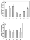
Analysis of wild-type and mutant HIV-1 virions. (A) Virus production in the culture medium after transfection of C33A cells with the proviral constructs. The results of three independent transfection experiments are summarized. Virus production for the wild-type (wt) pLAI construct was arbitrarily set at 1. (B) Packaging of HIV-1 RNA in virus particles. The packaging efficiency was measured by extension of the poly(A) primer on virion-isolated vRNA (Fig. 5, lanes 1 to 6). The results of three independent assays were quantitated, and the CA-p24 values were used to control for the amount of virions used per sample. Packaging was arbitrarily set at 1 for the wild-type virus.
FIG. 5.
FIG. 5.
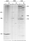
Reverse transcription assays with vRNA-tRNA complexes isolated from wild-type and mutant virions. The amount of vRNA was quantitated by DNA primer extension with the poly(A) primer, which produced a 104-nt product (lanes 1 to 6). Extension of the PBS-bound tRNA primer resulted in a 257-nt tRNA-cDNA product on the wild-type (wt) template (lanes 7 to 12). The products are shorter on the d1 and d1/2 template due to the d1 deletion in the U5 region. A major RT pause product with a length of 186 nt is visible on the wild-type template. The PBS-tRNA occupancy was determined by extension of the downstream AUG primer (lanes 13 to 18). When reverse transcription is blocked by the annealed tRNA primer, a 175-nt cDNA is produced. Free RNA templates will produce a full-length cDNA product of 374 nt. An additional stop product is observed on the 2L and d1 templates (lanes 13 and 16; stop). This stop is partially resolved on the d1 rev1 template (lane 17).
FIG. 6.
FIG. 6.
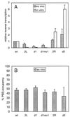
Relative reverse transcription activities of the wild-type and mutant templates. (A) Reverse transcription activity of virion-isolated vRNA-tRNA complexes as measured in Fig. 5 (ex vivo method). The results of three independent experiments were quantitated. There is a significant standard deviation for the d2 sample, which is due to the severe virus production defect of this mutant. The activity of the wild-type (wt) template was arbitrarily set at 1. For comparison, the results obtained previously with in vitro-assembled vRNA-tRNA complexes are included (8). (B) The tRNA occupancy of the PBS is shown for the wild-type and mutant vRNA genomes. The 374- and 175-nt products for three independent experiments were quantitated. The total of these products was arbitrarily set at 100%, and the percentage of 175-nt product is plotted.
FIG. 7.
FIG. 7.
Model for RNase H-mediated degradation of the vRNA template during reverse transcription. In the PBS occupancy assay, both the downstream AUG DNA primer and the tRNA primer are extended by RT. Extension of the tRNA primer yields a full-length tRNA-cDNA on the wild-type template (top) but not on the PAS-negative template (bottom). Extension of the tRNA primer triggers RNase H-mediated degradation of the RNA template (arrows and breaks in the processed RNA). The RNase H domain of HIV-1 RT is located 18 nt from the polymerase active site (24, 26). Thus, enhanced pausing of the RT enzyme near template position +154 on the PAS-negative template results in pronounced RNase H cleavage at position +172 (thick arrow). AUG-primed reverse transcription complexes that displace the PBS-bound tRNA primer stop at this position are shown. The HIV-1 RT enzyme covers approximately 8 nt upstream and 22 nt downstream of the polymerization site (34, 44), such that the reversion-based C150U mutation is within the RT complex.
FIG. 8.
FIG. 8.
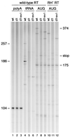
Reverse transcription with the wild-type and RNase H-negative RT enzyme. vRNA-tRNA complexes isolated from wild-type, d1, and d1 rev1 virions were used as templates for extension of the downstream AUG DNA primer (lanes 7 to 12). Reverse transcription was performed with the wild-type HIV-1 RT enzyme (lanes 7 to 9) and the RNase H-negative RT enzyme (RH RT; lanes 10 to 12). The major stop product (stop) on the d1 template that is observed upon extension of the AUG primer with wild-type RT (lane 8) is not observed with RNase H-negative RT (lane 11). AUG-primed reverse transcription with the RNase H-negative RT did not yield the full-length 374-nt product but produced several stop products. This is probably caused by the reduced processivity of the mutant RT enzyme. Control reactions were performed with the poly(A) primer (lanes 1 to 3) and the tRNA primer (lanes 4 to 6).
FIG. 9.
FIG. 9.
Model for HIV-1 reverse transcription. Shown is a schematic of the secondary structure in the PBS region of the HIV-1 RNA genome (black line) and the secondary structure of the tRNA3Lys molecule (orange line; AC, anticodon loop; D, D loop) that is used as a primer for reverse transcription. The tRNA primer with its 3"-terminal 18 nt anneals to the PBS for reverse transcription (PBS and anti-PBS are green), but an additional interaction between the PAS in the viral RNA and the anti-PAS in the tRNA primer (orange) is necessary for efficient initiation and elongation of reverse transcription.

Similar articles

Cited by

References

    1. Aiyar, A., D. Cobrinik, Z. Ge, H. J. Kung, and J. Leis. 1992. Interaction between retroviral U5 RNA and the TYC loop of the tRNATrp primer is required for efficient initiation of reverse transcription. J. Virol. 66:2464-2472. - PMC - PubMed
    1. Aiyar, A., Z. Ge, and J. Leis. 1994. A specific orientation of RNA secondary structures is required for initiation of reverse transcription. J. Virol. 68:611-618. - PMC - PubMed
    1. Arts, E. J., M. Ghosh, P. S. Jacques, B. Ehresmann, and S. F. J. Le Grice. 1996. Restoration of tRNALys,3-primed (−)-strand DNA synthesis to an HIV-1 reverse transcriptase mutant with extended tRNAs. J. Biol. Chem. 271:9054-9061. - PubMed
    1. Arts, E. J., and S. F. J. Le Grice. 1998. Interaction of retroviral reverse transcriptase with template-primer duplexes during replication. Prog. Nucleic Acid Res. Mol. Biol. 58:339-393. - PubMed
    1. Arts, E. J., S. R. Stetor, Y. Li, J. W. Rausch, K. J. Howard, B. Ehresmann, T. W. North, B. M. Wohrl, R. S. Goody, M. A. Wainberg, and S. F. J. Le Grice. 1996. Initiation of (−) strand DNA synthesis from tRNALys3 on lentiviral RNAs: implications of specific HIV-1 RNA-tRNALys3 interactions inhibiting primer utilization by retroviral reverse trancriptases. Proc. Natl. Acad. Sci. USA 93:10063-10068. - PMC - PubMed

Publication types

MeSH terms