. 2002 Mar;76(5):2340-9.
doi: 10.1128/jvi.76.5.2340-2349.2002.

Endocytosis of adeno-associated virus type 5 leads to accumulation of virus particles in the Golgi compartment

Affiliations

Endocytosis of adeno-associated virus type 5 leads to accumulation of virus particles in the Golgi compartment

Ursula Bantel-Schaal et al. J Virol. 2002 Mar.

Abstract

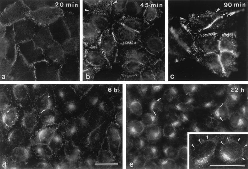
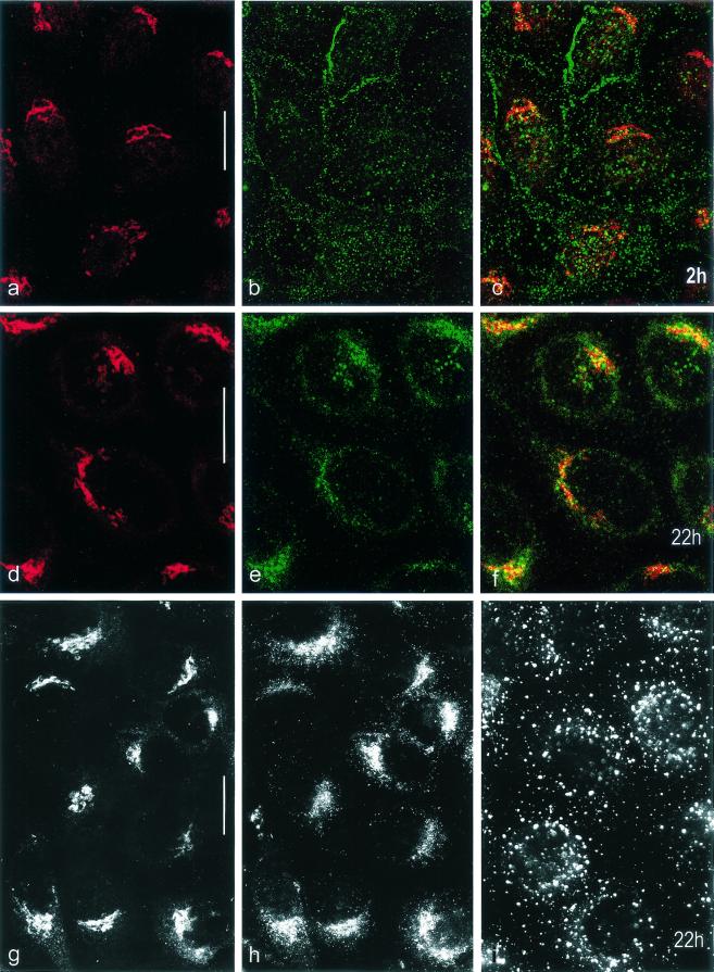
Among the adeno-associated virus (AAV) serotypes which are discussed as vectors for gene therapy AAV type 5 (AAV5) represents a candidate with unique advantages. To further our knowledge on AAV5-specific characteristics, we studied the entry pathway of wild-type virus in HeLa cells in the absence of helper virus by immunofluorescence and electron microscopy and by Western blot analysis. We found virus binding at the apical cell surface, especially at microvilli and, with increasing incubation time, virus accumulation at cell-cell boundaries. The different binding kinetics suggest different binding properties at apical versus lateral plasma membranes. Endocytosis of viruses was predominantly by clathrin-coated vesicles from both membrane domains; however, particles were also detected in noncoated pits. AAV5 particles were mainly routed to the Golgi area, where they could be detected within cisternae of the trans-Golgi network and within vesicles associated with cisternae and with the dictyosomal stacks of the Golgi apparatus. These data suggest that AAV5 makes use of endocytic routes that have hitherto not been described as pathways for virus entry.


Figures

FIG. 1.
FIG. 1.
(a) AAV5 particles negatively stained with 2% uranyl acetate in aqueous solution. Bar, 0.1 μm. (b to f) Electron-microscopic immunolocalization of AAV5 in HeLa cells. Cells were infected with AAV5 at 37°C and fixed with formaldehyde 90 min p.i. Immunoreactions were done using a monoclonal antibody specific for AAV5 capsids (21) and anti-mouse antibodies coupled to 5-nm gold particles for visualization. Since they were done prior to cell embedding, labeling could be detected only when AAV particles were accessible to the antibodies in the preembedding state. Thus only the laterally located particles in the flat section (b and c) are labeled with gold. Arrows, virus particles on (b and c) and between (d) microvilli, in a coated pit (e), in a coated vesicle (f), within an endosome (g), and within lysosomal structures (h and i). The concentration of AAV5 used in the experiments shown in panels e to i was 10-fold higher than that used in experiments shown in panels b to d. En, endosome; ER, rough endoplasmic reticulum; Ly, lysosomal structure; M, mitochondrion; PM, plasma membrane; Ri, ribosomes; Bars, 0.1 μm (f; applies to panels b to f) and 0.2 μm (i; applies to panels g to i).
FIG. 2.
FIG. 2.
Immunofluorescence microscopy of HeLa cells after incubation with AAV5. HeLa cells were cultured in the presence of AAV5 for the indicated periods of time and were then fixed with methanol-acetone before incubation with antibodies. Removal of the inoculum was after 90 min (c) and was followed by three washes before addition of fresh medium. Arrowheads (b and c), viruses at apical cell surfaces and in endocytic structures; arrows (d and e), fluorescent aggregates in juxtanuclear position. Inset (e), additional faint positive reactions at the nuclear envelope (arrowheads). Bars (d and inset), 30 μm (the bar in panel d applies to panels a to e [except for the inset]).
FIG. 3.
FIG. 3.
Distribution of fluorescence signals on AAV5-infected HeLa cells. HeLa cells were infected with AAV5 for 2 (a to c) and 22 h (d to i). At the times indicated cells were fixed with methanol-acetone (a to f) or with formaldehyde (g to i). Double labeling (confocal laser scanning microscopy; optical sections, 0.6 μm thick) (a to f) was done using primary antibodies specific for Golgi ManII (a and d) and for AAV5 capsids (b and e). The respective merged images are shown in panels c and f. The images in panels g to i show that the signal distributions for antibodies to ManII (g) and AAV5 capsids (h) are different from that for the antibody to EEA1 (endosomes) (i) at 22 h p.i. Formaldehyde fixation was used (g to i) to verify the results obtained with methanol-acetone fixation. Bars, 20 μm.
FIG. 4.
FIG. 4.
Electron micrographs of HeLa cells demonstrating binding and early steps of internalization. HeLa cells were infected with AAV5 for 20 (a to c), 45 (d to i), and 90 min (k to o). AAV5 particles are seen at apical (a to i) and lateral (k to o) cell surfaces (small arrows). Arrowheads (b), cross-sectioned microtubules as an internal size marker. Internalization of viruses by coated vesicles takes place from apical (c to g) and lateral (m to o) cell surfaces (large arrows). Tu (f), example of particles in tubular structures that are likely to represent early endosomes. Aggregated viruses at lateral surfaces are demonstrated in cross (k, m, and n) and oblique (l, brackets) sections. In rare situations internalization via noncoated pits and vesicles was also seen (h and i). CP, coated pit; ER, endoplasmic reticulum; MV, microvilli; D, desmosome. Bars, 0.2 μm (a, b, and k; same magnification for panels k to o) and 0.1 μm (c to i).
FIG. 5.
FIG. 5.
Electron micrographs of HeLa cells infected with AAV5 particles taken 3 (a and b), 6 (c, e, h, and i), and 22 h (d, f, and g) p.i. Viruses (small arrows) were found in clathrin-coated vesicles (CL; see, e.g., panel a), in tubular (large arrows; b and e) and vesicular structures (large arrows; g), and in noncoated vesicles in the vicinity of dilated Golgi cisternae and stacks of the Golgi apparatus (GA; large arrows). Examples of cisternae of the TGN containing AAV5 particles are given in panels c, d, g, h, and i. Co, coatomer-coated vesicles. Bars, 0.2 μm.
FIG. 6.
FIG. 6.
Western blot analysis of HeLa cell proteins after incubation with AAV5 particles. After the indicated periods of time (lanes 3 to 8) cells were washed and homogenized. Equal amounts of proteins (cells) were loaded onto the gel. After 90 min the inoculum was replaced by fresh virus-free culture medium (lanes 6 to 8). Lane 1, calculated virus concentration used in infection. A 10-fold concentration (lane 9) was used to demonstrate that the antibody recognizes all three capsid proteins. Lane 2, mock-infected cells.
FIG. 7.
FIG. 7.
Scheme of proposed endocytic pathway for AAV5 into HeLa cells. CA, noncoated pit; CP, coated pit; CV, coated vesicle; En, endosome; MV, microvilli; NP, nuclear pore; D, desmosome.

Similar articles

Cited by

References

    1. Balague, C., M. Kalla, and W. W. Zhang. 1997. Adeno-associated virus Rep78 protein and terminal repeats enhance integration of DNA sequences into the cellular genome. J. Virol. 71:3299-3306. - PMC - PubMed
    1. Bantel-Schaal, U., H. Delius, R. Schmidt, and H. zur Hausen. 1999. Human adeno-associated virus type 5 is only distantly related to other known primate helper-dependent parvoviruses. J. Virol. 73:939-947. - PMC - PubMed
    1. Bantel-Schaal, U., and H. zur Hausen. 1984. Characterization of the DNA of a defective parvovirus isolated from a genital site. Virology 134:52-63. - PubMed
    1. Bantel-Schaal, U., and H. zur Hausen. 1988. Adeno-associated viruses inhibit SV40 DNA amplification and replication of herpes simplex virus in SV40-transformed hamster cells. Virology 164:64-74. - PubMed
    1. Bartlett, J. S., R. Wilcher, and R. J. Samulski. 2000. Infectious entry pathway of adeno-associated virus and adeno-associated virus vectors. J. Virol. 74:2777-2785. - PMC - PubMed

LinkOut - more resources