. 2002 Mar;76(5):2393-402.
doi: 10.1128/jvi.76.5.2393-2402.2002.

The capsid of infectious bursal disease virus contains several small peptides arising from the maturation process of pVP2

Affiliations

The capsid of infectious bursal disease virus contains several small peptides arising from the maturation process of pVP2

Bruno Da Costa et al. J Virol. 2002 Mar.

Abstract

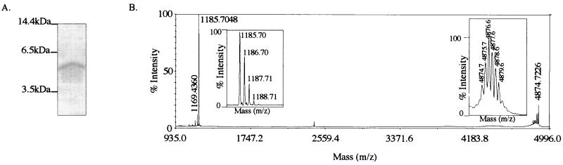
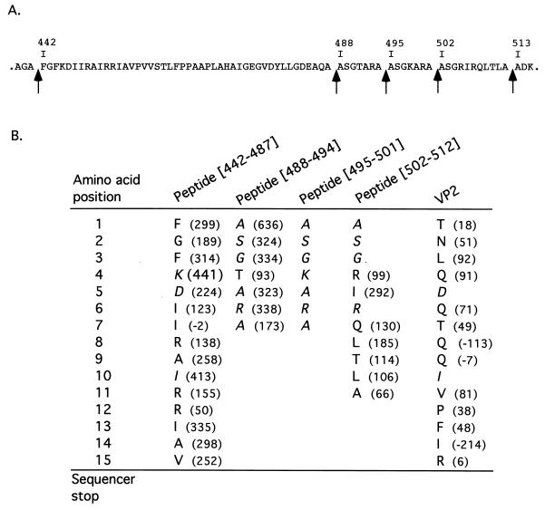
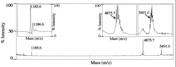
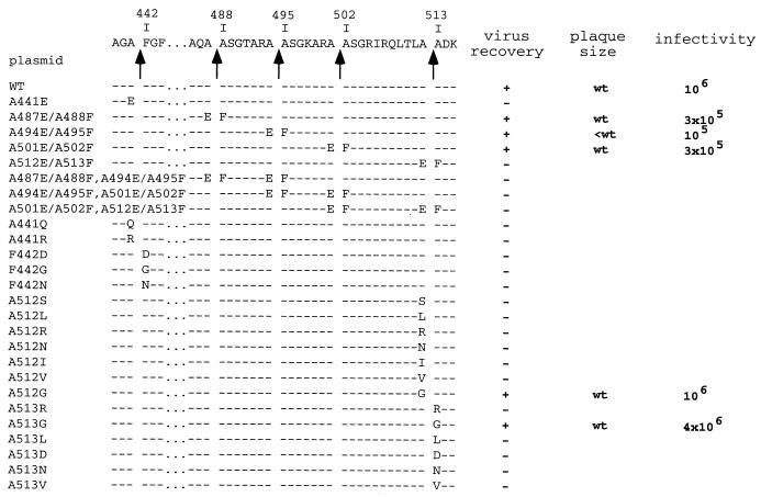
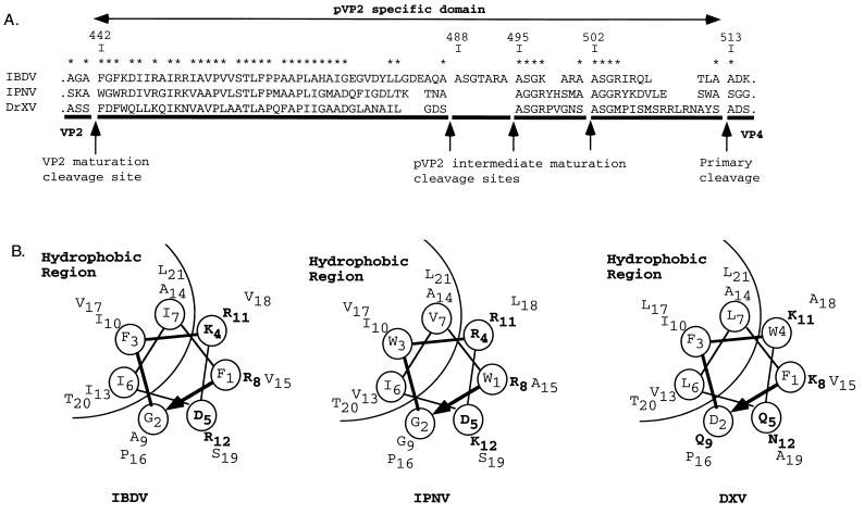
The capsid proteins VP2 and VP3 of infectious bursal disease virus, a birnavirus, are derived from the processing of a large polyprotein: NH2-pVP2-VP4-VP3-COOH. Although the primary cleavage sites at the pVP2-VP4 and VP4-VP3 junctions have been identified, the proteolytic cascade involved in the processing of this polyprotein is not yet fully understood, particularly the maturation of pVP2. By using different approaches, we showed that the processing of pVP2 (residues 1 to 512) generated VP2 and four small peptides (residues 442 to 487, 488 to 494, 495 to 501, and 502 to 512). We also showed that in addition to VP2, at least three of these peptides (residues 442 to 487, 488 to 494, and 502 to 512) were associated with the viral particles. The importance of the small peptides in the virus cycle was assessed by reverse genetics. Our results showed that the mutants lacking the two smaller peptides were viable, although the virus growth was affected. In contrast, deletions of the domain 442 to 487 or 502 to 512 did not allow virus recovery. Several amino acids of the peptide 502 to 512 appeared essential for virus viability. Substitutions of the P1 and/or P1" position were engineered at each of the cleavage sites (P1-P1": 441-442, 487-488, 494-495, 501-502, and 512-513). Most substitutions at the pVP2-VP4 junction (512-513) and at the final VP2 maturation cleavage site (441-442) were lethal. Mutations of intermediate cleavage sites (487-488, 494-495, and 501-502) led to viable viruses showing different but efficient pVP2 processing. Our data suggested that while peptides 488 to 494 and 495 to 501 play an accessory role, peptides 442 to 487 and 502 to 512 have an unknown but important function within the virus cycle.


Figures

FIG. 1.
FIG. 1.
Characterization of peptides present in IBDV particles. (A) SDS-PAGE (16% polyacrylamide) analysis of purified virus. A peptide of about 5 kDa was identified and excised from the gel for N-terminal sequence analysis. Its N terminus was mapped at residue 442. Molecular mass markers are indicated on the left. (B) Mass spectrometry analysis of IBDV particles. Two main signals were identified on the mass/charge window ranging from 935 to 4,996. Magnified signals showing the isotopic pattern are inserted. The [M + H]+ values for the peptides are 1,185.7 and 4,874.7 Da, respectively.
FIG. 2.
FIG. 2.
N-terminal sequencing of the purified virus. (A) Sequence of the pVP2-specific domain from amino acids 442 to 512. The single-letter code is used to indicate amino acids. Arrows indicate the cleavage sites. (B) The nature of the amino acids and the amount (differential picomoles) revealed at each Edman degradation cycle (amino acid position) are indicated. Sequences of the peptides (and of the mature VP2) have been aligned on the identified residues. Residues which are potentially present in more than one peptide are indicated in italics.
FIG. 3.
FIG. 3.
Mass spectrometry analysis of IBDV VLPs. Three main signals were identified in the mass/charge window. Magnified signals showing the isotopic pattern are inserted. The [M + H]+ values for the peptides are 1,185.6, 4,875.7 and 5,491.0 Da, respectively.
FIG. 4.
FIG. 4.
Comparison of the electrophoretic mobilities of the different forms of VP2. Mock-infected (−) or IBDV-infected [IBDV(CT)] cells were 35S labeled and subjected to immunoprecipitation with an anti-VP2 monoclonal antibody. Denatured immune complexes were run side by side on an SDS-PAGE gel with an in vitro translation product of the mutated IBDA polyprotein having a stop codon at position 442 (VP2/1-441). The gel was subjected to autoradiography. Molecular mass markers are indicated on the left.
FIG. 5.
FIG. 5.
Virus recovery for the pVP2 cleavage site mutants. The amino acid sequence of the pVP2-specific domain and the different cleavage sites (arrows) are indicated at the top of the figure. The mutated amino acid(s) is indicated for each construct in the single-letter code. Depending on the virus recovery results, the plasmids are indicated by a + (recovery) or − (nonrecovery). The relative plaque size at 48 h after infection of confluent LMH cell monolayers is indicated. wt, wild-type size; small, smaller than wild-type size. Rescued viruses were amplified on LSCC-BK3 cells, and virus titers were determined 3 days postinfection on LMH cells.
FIG. 6.
FIG. 6.
Phenotypes of several mutants. (A) Typical aspect of plaques formed by the indicated mutants. The dilutions used for the assay are indicated. (B) Analysis of the pVP2 processing in cells infected with the indicated mutants. Left lanes show VP2 immunoprecipitations of the cell extracts; right lanes show VP2 immunoprecipitations of the cell supernatants. In the middle is shown mature VP2 (VP2/1-441) expressed in vitro. IBDV (CT) indicates the results of a regular (nonrescued) infection. Lane A512E-A513F, no rescued virus with the corresponding plasmid.
FIG. 7.
FIG. 7.
In vitro processing of different P1 or P1" mutants. The autoradiograph shows the results obtained with pT7-IBDA-HDR (wt) and a set of P1 or P1" mutants produced in a rabbit reticulocyte expression system. Expression products were analyzed by SDS-PAGE (10% polyacrylamide), and the gel was processed for autoradiography. The positions of the viral polypeptides are indicated on the right.
FIG. 8.
FIG. 8.
Virus recovery of the pVP2 mutants. The amino acid sequence of the pVP2-specific domain and the different cleavage sites (arrows) are indicated at the top of the figure. Deleted amino acids are indicated by an asterisk, and substitutions are indicated for each plasmid in the single-letter code. Depending on the virus recovery results, the plasmids are indicated by + (recovery) or − (nonrecovery). Plaque size and virus production were analyzed as described in the legend to Fig. 5.
FIG. 9.
FIG. 9.
The pVP2-specific domain of different birnavirus polyproteins. (A) Sequence alignment of the IBDV, infectious pancreatic necrosis virus (IPNV), and Drosophila X virus (DXV) pVP2-specific domains. The alignment is anchored to the multiple cleavage sites identified on IBDV or proposed for infectious pancreatic necrosis virus (18) and to the cleavage site at the pVP2-VP4 junction of Drosophila X virus (8). Stars indicate residues common to two or more sequences. (B) Helical-wheel representation of the putative amphipathic α-helices of the N-terminal domains of the [442-487] IBDV peptide and its infectious pancreatic necrosis virus and Drosophila X virus homologs.

References

    1. Azad, A. A., M. N. Jagadish, M. A. Brown, and P. J. Hudson. 1987. Deletion mapping and expression in Escherichia coli of the large genomic segment of a birnavirus. Virology 161:145-152. - PubMed
    1. Biacchesi, S., Y. X. Yu, M. Bearzotti, C. Tafalla, M. Fernandez-Alonso, and M. Bremont. 2000. Rescue of synthetic salmonid rhabdovirus minigenomes. J. Gen. Virol. 81:1941-1945. - PubMed
    1. Bong, D. T., C. Steinem, A. Janshoff, J. E. Johnson, and M. R. Ghadiri. 1999. A highly membrane-active peptide in Flock House virus: implications for the mechanism of nodavirus infection. Chem. Biol. 6:473-481. - PubMed
    1. Bong, D. T., A. Janshoff, C. Steinem, and M. R. Ghadiri. 2000. Membrane partitioning of the cleavage peptide in flock house virus. Biophys. J. 78:839-845. - PMC - PubMed
    1. Boot, H. J., A. A. H. M. ter Huurne, B. P. H. Peeters, and A. L. J. Gielkens. 1999. Efficient rescue of infectious bursal disease virus from cloned cDNA: evidence for involvement of the 3"-terminal sequence in genome replication. Virology 265:330-341. - PubMed

MeSH terms

Substances

LinkOut - more resources