. 2002 Mar;76(5):2518-28.
doi: 10.1128/jvi.76.5.2518-2528.2002.

Ebola virus glycoproteins induce global surface protein down-modulation and loss of cell adherence

Affiliations

Ebola virus glycoproteins induce global surface protein down-modulation and loss of cell adherence

Graham Simmons et al. J Virol. 2002 Mar.

Abstract

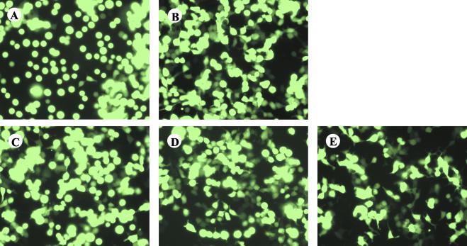
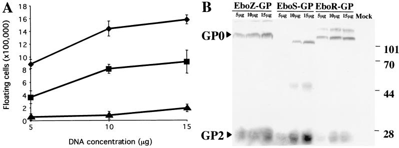
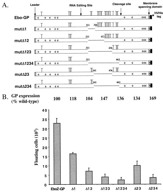
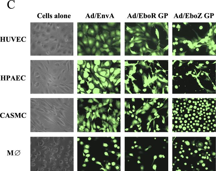
The Ebola virus envelope glycoprotein (GP) derived from the pathogenic Zaire subtype mediates cell rounding and detachment from the extracellular matrix in 293T cells. In this study we provide evidence that GPs from the other pathogenic subtypes, Sudan and Côte d'Ivoire, as well as from Reston, a strain thought to be nonpathogenic in humans, also induced cell rounding, albeit at lower levels than Zaire GP. Sequential removal of regions of potential O-linked glycosylation at the C terminus of GP1 led to a step-wise reduction in cell detachment without obviously affecting GP function, suggesting that such modifications are involved in inducing the detachment phenotype. While causing cell rounding and detachment in 293T cells, Ebola virus GP did not cause an increase in cell death. Indeed, following transient expression of GP, cells were able to readhere and continue to divide. Also, the rounding effect was not limited to 293T cells. Replication-deficient adenovirus vectors expressing Ebola virus GP induced the loss of cell adhesion in a range of cell lines and primary cell types, including those with proposed relevance to Ebola virus infection in vivo, such as endothelial cells and macrophages. In both transfected 293T and adenovirus-infected Vero cells, a reduction in cell surface expression of adhesion molecules such as integrin beta1 concurrent with the loss of cell adhesion was observed. A number of other cell surface molecules, however, including major histocompatibility complex class I and the epidermal growth factor receptor, were also down-modulated, suggesting a global mechanism for surface molecule down-regulation.


Figures

FIG. 1.
FIG. 1.
Ebola virus GP expression induces cell rounding. 293T cells were transfected with 20 μg of pAD-TrackCMV containing the coding sequence for EboZ GP (A), EboS GP (B), EboR GP (C), EboC-GP (D), or ASLV-A EnvA (E). GFP expression was observed at 48 h posttransfection using a Nikon TE300 inverted microscope. Fields represent findings from multiple experiments.
FIG. 2.
FIG. 2.
The cell rounding phenotype correlates with the level of GP expression. (A) 293T cells were transfected with 5, 10, or 15 μg of pcDNA6/V5-His containing the coding sequence for EboZ GP (♦), EboS GP (▪), or EboR GP (▴). The floating cells in the cultures were counted 48 h posttransfection, and results are presented as numbers of floating cells above background levels (8 × 10⁴ cells), calculated from mock-transfected cells. (B) Lysates from cells harvested 48 h posttransfection were subjected to SDS-4 to 15% PAGE, transferred to nitrocellulose, and immunoblotted for the presence of the C-terminal V5 epitope on both the uncleaved precursor (GP0) and processed GP2 using a mouse anti-V5 monoclonal antibody followed by I125-labeled protein A. Data are representative of three independent experiments.
FIG. 3.
FIG. 3.
Cell rounding is dependent on O-linked glycosylation-rich domains in GP1. (A) Serial deletions in the C-terminal mucin-like domain of GP1 were made by overlapping extension PCR. Potential O-linked glycosylations (T) were predicted using NetOGlyc, version 2.0. (B) 293T cells were transfected with 30 μg of pcDNA6/V5-His containing the coding sequences for EboZ GP or EboZ GP mucin domain mutants. Floating cells were counted, and values are numbers of floating cells above background levels (2.4 × 10⁵ cells), calculated from mock-transfected cells. Cell lysates from 5 × 10⁶ cells were subjected to SDS-4 to 15% PAGE, and mature-GP2 levels were quantified by phosphorimager analysis following immunoblotting for the presence of the V5 epitope using a mouse anti-V5 monoclonal antibody followed by I125-labeled protein A. GP2 levels are presented as a percentage of EboZ GP2 wild-type expression. Data are representative of three independent experiments.
FIG. 4.
FIG. 4.
Ebola virus GP transduction of human and nonhuman cell lines and primary cell types using adenovirus vectors. (A) Human cell lines U87, 293H, and PMA-pretreated U937 were transduced with the minimum inoculum of adenovirus vectors expressing ASLV-A EnvA, EboR GP, or EboZ GP required to give the highest achievable level of GFP-positive cells (MOIs of 30, 10, and 50, respectively). Cells were monitored at regular intervals for evidence of cell rounding, and representative photographs were taken at 48 h posttransduction. (B) Adenovirus vectors expressing ASLV-A EnvA, EboR GP, or EboZ GP were used to transduce a simian cell line, Vero (MOI, 10), a cat kidney cell line, CCC (MOI, 10), baby hamster kidney cells (BHK; MOI, 50), and a murine cell line expressing the human coxsackievirus/adenovirus receptor, MC57/hCAR (MOI, 50). Cells were monitored for rounding as described for panel A. AGM, African green monkey. (C) Adenovirus vectors expressing ASLV-A EnvA, EboR GP, or EboZ GP were used to transduce low-passage primary HUVEC, HPAEC, CASMC as well as day 8 human primary blood monocyte-derived macrophages (MDM). Similar results were obtained for MDM from three separate donors. An MOI of 10 was used for all primary cell types, apart from MDM, where an MOI of 50 was required. Cells were monitored for rounding as described for panel A. In all cases, control vectors expressing just GFP gave results indistinguishable from those for EnvA (data not shown). M⊘, macrophage.
FIG. 4.
FIG. 4.
Ebola virus GP transduction of human and nonhuman cell lines and primary cell types using adenovirus vectors. (A) Human cell lines U87, 293H, and PMA-pretreated U937 were transduced with the minimum inoculum of adenovirus vectors expressing ASLV-A EnvA, EboR GP, or EboZ GP required to give the highest achievable level of GFP-positive cells (MOIs of 30, 10, and 50, respectively). Cells were monitored at regular intervals for evidence of cell rounding, and representative photographs were taken at 48 h posttransduction. (B) Adenovirus vectors expressing ASLV-A EnvA, EboR GP, or EboZ GP were used to transduce a simian cell line, Vero (MOI, 10), a cat kidney cell line, CCC (MOI, 10), baby hamster kidney cells (BHK; MOI, 50), and a murine cell line expressing the human coxsackievirus/adenovirus receptor, MC57/hCAR (MOI, 50). Cells were monitored for rounding as described for panel A. AGM, African green monkey. (C) Adenovirus vectors expressing ASLV-A EnvA, EboR GP, or EboZ GP were used to transduce low-passage primary HUVEC, HPAEC, CASMC as well as day 8 human primary blood monocyte-derived macrophages (MDM). Similar results were obtained for MDM from three separate donors. An MOI of 10 was used for all primary cell types, apart from MDM, where an MOI of 50 was required. Cells were monitored for rounding as described for panel A. In all cases, control vectors expressing just GFP gave results indistinguishable from those for EnvA (data not shown). M⊘, macrophage.
FIG. 5.
FIG. 5.
Ebola virus GP does not induce cell death. Floating cells (3 × 10⁵ per well) from 293T cells 48 h posttransfection were replated into fresh six-well plates. At 24-h intervals floating cells were counted (•) and then pooled with versine-detached adherent cells, and viable (♦) and nonviable (▪) cell numbers were estimated using trypan blue exclusion. Cells were then adhered to poly-d-lysine-coated plates, and the percentage of Ebola virus GP-positive cells (▴) was quantified using indirect immunofluorescence staining with KZ52. Values represent the geometric means of triplicate samples ± standard errors. Data are representative of three independent experiments.
FIG. 6.
FIG. 6.
Expression of Ebola virus GP induces down-modulation of cell surface molecules. (A) At 48 h after transfection with pCB6-EboZ-GP, -EboR-GP, or -ASLV-A-EnvA, 293T cells were detached from the tissue culture plates, pooled with already-floating cells, and incubated with monoclonal antibodies directed against a number of integrin subunits as well as other common cell surface markers. Flow-cytometric analysis was then performed using secondary antibodies conjugated with fluorescein. Results are presented percentages of the mean channel fluorescence (MCF) compared to that for EnvA-transfected cells. Data are representative of multiple independent experiments. (B) Vero cells transduced at an MOI of 10 with Ad/EnvA Ad/EboR-GP, or EboZ GP were collected at 48 h postinfection. Cells were incubated with monoclonal antibodies to an isotype control, β1-integrin, αv-integrin, MHC-I, or EGFR and a secondary antibody conjugated to Cy5 and assayed by flow cytometry for GFP expression (FL1; y axis) or cell surface markers (FL4; x axis). Dotted lines, estimated MCF of expression for mock-transduced cells (C) HUVEC transduced at an MOI of 10 with Ad/EboZ-GP (solid line), Ad/EboR-GP (dashed line), or Ad/EnvA (dotted line) were collected 24 h postinfection. Cells were incubated with monoclonal antibodies to isotype controls (shaded histograms), β1-integrin, αv-integrin, MHC-I, or PECAM and a secondary antibody conjugated to Cy5 and assayed by flow cytometry. In HUVEC, unlike HPAEC, which showed up-regulation of some markers in response to adenovirus infection, there was no difference between nontransduced and Ad/EnvA-transduced cells for any of the markers (data not shown). Data are representative of three independent experiments.
FIG. 6.
FIG. 6.
Expression of Ebola virus GP induces down-modulation of cell surface molecules. (A) At 48 h after transfection with pCB6-EboZ-GP, -EboR-GP, or -ASLV-A-EnvA, 293T cells were detached from the tissue culture plates, pooled with already-floating cells, and incubated with monoclonal antibodies directed against a number of integrin subunits as well as other common cell surface markers. Flow-cytometric analysis was then performed using secondary antibodies conjugated with fluorescein. Results are presented percentages of the mean channel fluorescence (MCF) compared to that for EnvA-transfected cells. Data are representative of multiple independent experiments. (B) Vero cells transduced at an MOI of 10 with Ad/EnvA Ad/EboR-GP, or EboZ GP were collected at 48 h postinfection. Cells were incubated with monoclonal antibodies to an isotype control, β1-integrin, αv-integrin, MHC-I, or EGFR and a secondary antibody conjugated to Cy5 and assayed by flow cytometry for GFP expression (FL1; y axis) or cell surface markers (FL4; x axis). Dotted lines, estimated MCF of expression for mock-transduced cells (C) HUVEC transduced at an MOI of 10 with Ad/EboZ-GP (solid line), Ad/EboR-GP (dashed line), or Ad/EnvA (dotted line) were collected 24 h postinfection. Cells were incubated with monoclonal antibodies to isotype controls (shaded histograms), β1-integrin, αv-integrin, MHC-I, or PECAM and a secondary antibody conjugated to Cy5 and assayed by flow cytometry. In HUVEC, unlike HPAEC, which showed up-regulation of some markers in response to adenovirus infection, there was no difference between nontransduced and Ad/EnvA-transduced cells for any of the markers (data not shown). Data are representative of three independent experiments.

References

    1. Alon, R., P. D. Kassner, M. W. Carr, E. B. Finger, M. E. Hemler, and T. A. Springer. 1995. The integrin VLA-4 supports tethering and rolling in flow on VCAM-1. J. Cell Biol. 128:1243-1253. - PMC - PubMed
    1. Anonymous. 1990. Update: filovirus infection in animal handlers. Morbid. Mortal. Wkly. Rep. 39:221. - PubMed
    1. Baize, S., E. M. Leroy, M. C. Georges-Courbot, M. Capron, J. Lansoud-Soukate, P. Debre, S. P. Fisher-Hoch, J. B. McCormick, and A. J. Georges. 1999. Defective humoral responses and extensive intravascular apoptosis are associated with fatal outcome in Ebola virus-infected patients. Nat. Med. 5:423-426. - PubMed
    1. Balter, M. 2000. On the trail of Ebola and Marburg viruses. Science 290:923-924. - PubMed
    1. Baskerville, A., S. P. Fisher-Hoch, G. H. Neild, and A. B. Dowsett. 1985. Ultrastructural pathology of experimental Ebola haemorrhagic fever virus infection. J. Pathol. 147:199-209. - PubMed

Publication types

Substances