Review
. 2002 Feb;32(2):65-74.
doi: 10.2519/jospt.2002.32.2.65.

Muscular, skeletal, and neural adaptations following spinal cord injury

Affiliations
Review

Muscular, skeletal, and neural adaptations following spinal cord injury

Richard K Shields. J Orthop Sports Phys Ther. 2002 Feb.

Abstract

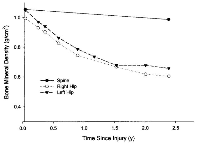
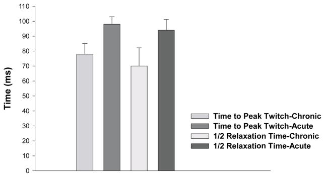
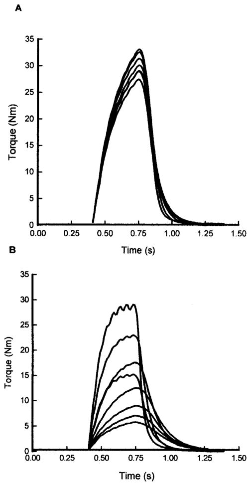
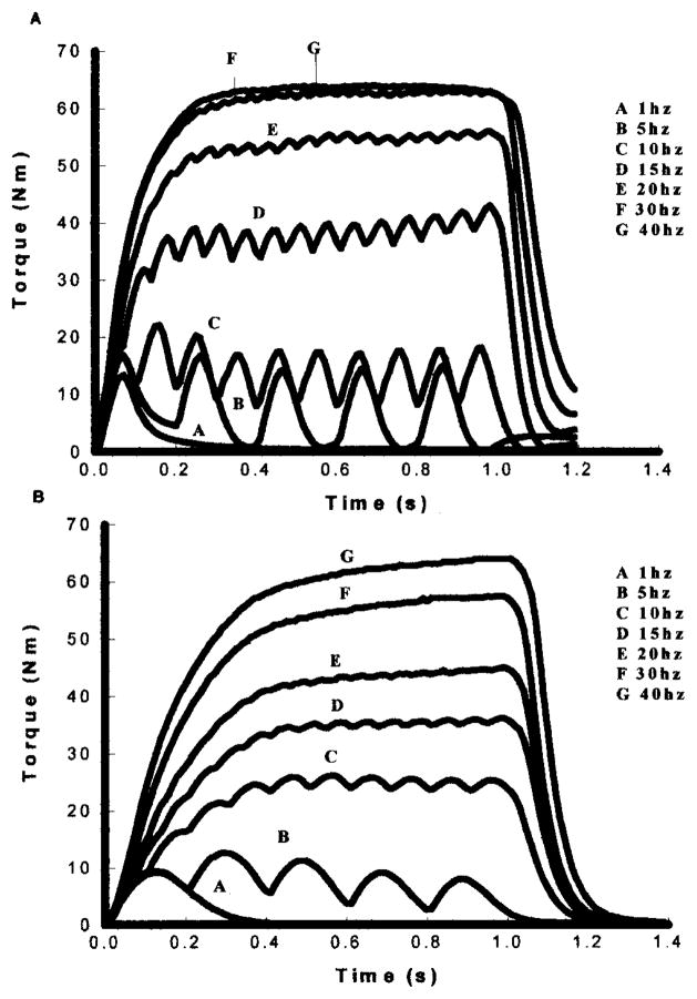
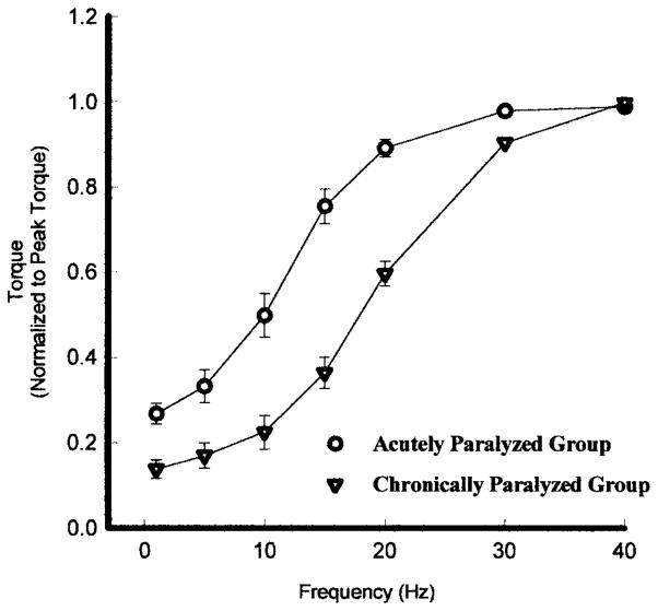
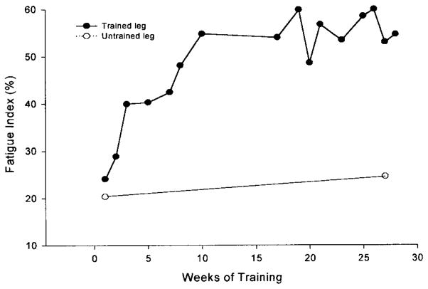
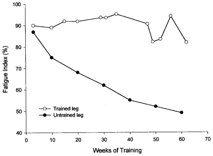
Spinal cord injury is associated with adaptations to the muscular, skeletal, and spinal systems. Experimental data are lacking regarding the extent to which rehabilitative methods may influence these adaptations. An understanding of the plasticity of the muscular, skeletal, and spinal systems after paralysis may be important as new rehabilitative technologies emerge in the 21st century. Moreover, individuals injured today may become poor candidates for future scientific advancements (cure) if their neuromusculoskeletal systems are irreversibly impaired. The primary purpose of this paper is to explore the physiological properties of skeletal muscle as a result of spinal cord injury; secondarily, to consider associated changes at the skeletal and spinal levels. Muscular adaptations include a transformation to faster myosin, increased contractile speeds, shift to the right on the torque-frequency curve, increased fatigue, and enhanced doublet potentiation. These muscular adaptations may be prevented in individuals with acute paralysis and partially reversed in individuals with chronic paralysis. Moreover, the muscular changes may be coordinated with motor unit and spinal circuitry adaptations. Concurrently, skeletal adaptations, as measured by bone mineral density, show extensive loss within the first six months after paralysis. The underlying science governing neuromusculoskeletal adaptations after paralysis will help guide professionals as new rehabilitation strategies evolve in the future.


Figures

FIGURE 1
FIGURE 1
The bone mineral density (BMD) of the right hip (○), left hip (▼), and the lumbar spine (●) from the onset of injury to 2.5 years following complete quadriplegia. Notice the significant loss of bone density of the hips while the lumbar spine remains normal.
FIGURE 2
FIGURE 2
Chronically paralyzed rectus femoris muscle (A) and a normal rectus femoris muscle (B) stained for the myosin ATPase enzyme with a preincubation at pH 9.4 (1), pH 4.2 (2), and pH 4.6 (3). The oxidative enzyme NADH–TR was stained for in 4. Notice that the paralyzed muscle (A) appears as a homogenous group of type II fibers while the nonparalyzed muscle (B) shows the normal mixed distribution of type I and type II fibers.
FIGURE 3
FIGURE 3
A transmission electron micrograph of the chronically paralyzed soleus muscle. From one dark band (z–line) to the next represents one sarcomere. A series of micrographs revealed that the z–lines were, on average, 20 nanometers narrower in chronically paralyzed muscle. Narrow z-lines have previously been described for fast muscle. Magnification = 36,000x. Bar = 1 μm.
FIGURE 4
FIGURE 4
The mean time to peak twitch and mean half-relaxation time for the chronic and acute paralyzed soleus muscle. Error bars are standard deviations.
FIGURE 5
FIGURE 5
The torque curve sampled every 30 seconds during a 20-Hz fatigue protocol (every second for 330 ms) for an individual with acute (A) and chronic (B) paralysis. Notice the extensive fatigue and contractile speed slowing at the end of the fatigue protocol in the chronically paralyzed soleus.
FIGURE 6
FIGURE 6
The torque curves obtained with electrical stimulation at 1, 5, 10, 15, 20, 30, and 40 Hz for an individual with chronic paralysis (A) and an individual paralyzed for less than one week (B). Notice the difference in contractile speeds and fusion properties between the acute and chronically paralyzed soleus.
FIGURE 7
FIGURE 7
The average torque–frequency curve, normalized for each subjects maximal torque, for an acutely paralyzed group (○) and a chronically paralyzed group (▽). Because of the faster contractile speeds the chronically paralyzed group curve is shifted to the fight. Error bars are standard deviations.
FIGURE 8
FIGURE 8
Chronic Paralysis—The fatigue index (final torque/initial torque) from a repetitive stimulation protocol (15 Hz applied each second for 330 ms) for the trained and untrained soleus in an individual with chronic paralysis. Notice that after ten weeks of training the chronic soleus muscle showed more than 100% increase in its endurance while control (untrained) leg changed minimally.
FIGURE 9
FIGURE 9
Acute Paralysis—The fatigue index (final torque/initial torque) from a repetitive stimulation protocol (15 Hz applied each second for 330 ms) for a trained leg and untrained leg for an individual that began training one leg within two weeks of spinal cord injury. Notice that the trained leg kept the normal soleus fatigue resistance properties after one year of spinal cord injury.

References

    1. Allen DG, Lee JA, Westerblad H. Intracellular calcium and tension during fatigue in isolated single muscle fibres from Xenopus laevis. Physiol. 1989;415:433–458. - PMC - PubMed
    1. Bevan L, Laouris Y, Reinking RM, Stuart DG. The effect of the stimulation pattern on the fatigue of single motor units in adult cats. J Physiol. 1992;449:85–108. - PMC - PubMed
    1. Biering-Sorensen F, Bohr H, Schaadt O. Bone mineral content of the lumbar spine and lower extremities years after spinal cord lesion. Paraplegia. 1988;26:293–301. - PubMed
    1. Biering-Sorensen F, Bohr HH, Schaadt OP. Longitudinal study of bone mineral content in the lumbar spine, the forearm and the lower extremities after spinal cord injury. Eur J Clin Invest. 1990;20:330–335. - PubMed
    1. Binder-Macleod ST, Barker CB. Use of a catchlike property of human skeletal muscle to reduce fatigue. Muscle Nerve. 1991;14:850–857. - PubMed

Publication types

MeSH terms