. 2002 Feb;160(2):655-66.
doi: 10.1016/S0002-9440(10)64885-7.

Axonal injury in cerebral malaria

Affiliations

Axonal injury in cerebral malaria

Isabelle M Medana et al. Am J Pathol. 2002 Feb.

Abstract

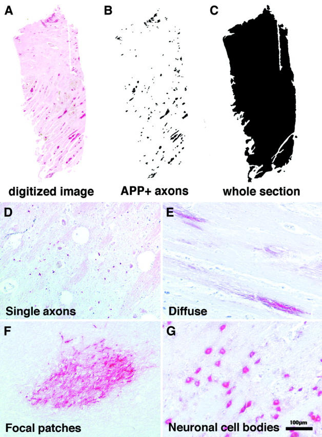
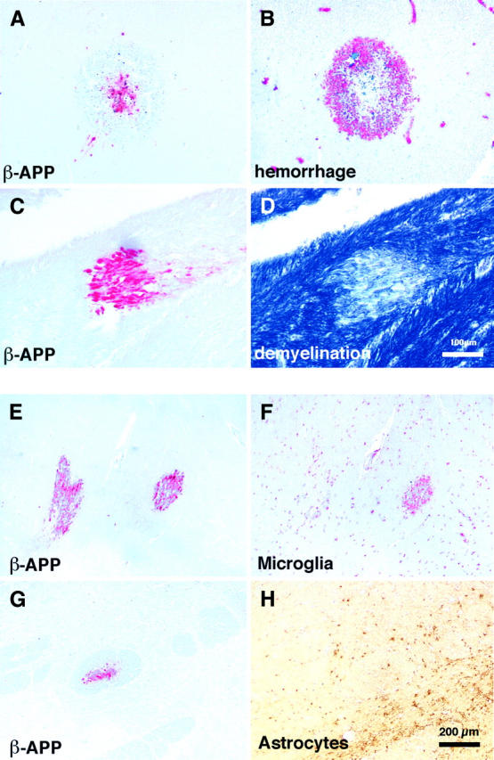
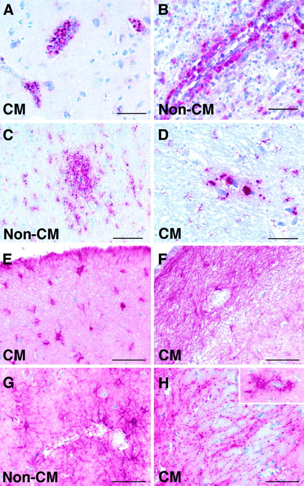
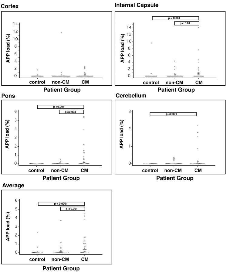
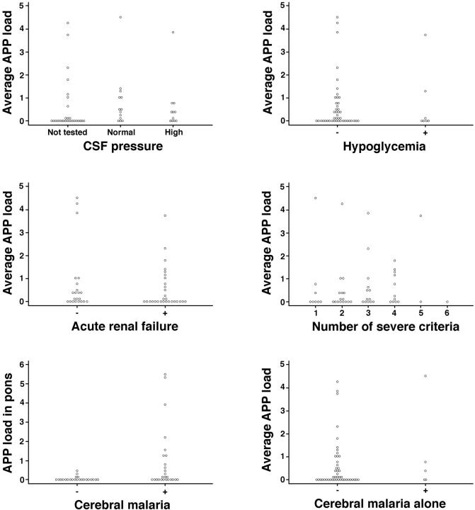
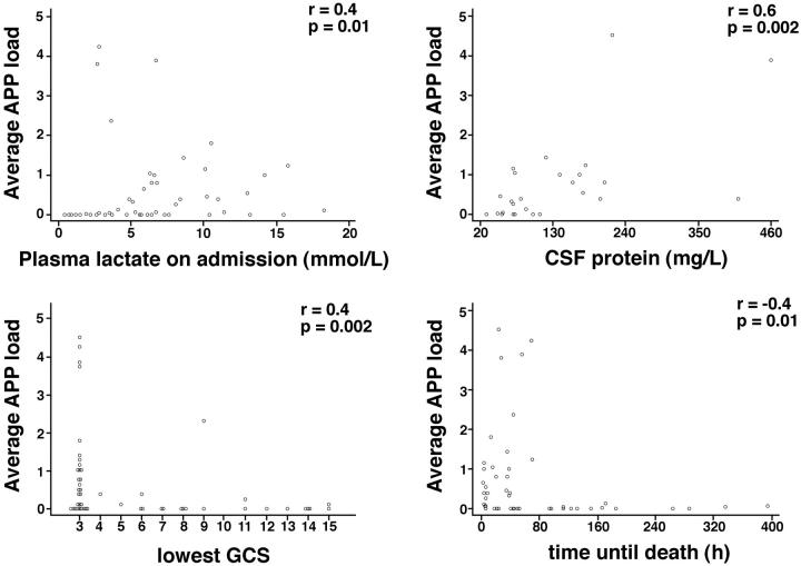
Impairment of consciousness and other signs of cerebral dysfunction are common complications of severe Plasmodium falciparum malaria. Although the majority of patients make a complete recovery a significant minority, particularly children, have sequelae. The pathological process by which P. falciparum malaria induces severe but usually reversible neurological complications has not been elucidated. Impairment of transport within nerve fibers could induce neurological dysfunction and may have the potential either to resolve or to progress to irreversible damage. Beta-amyloid precursor protein (beta-APP) immunocytochemistry, quantified using digital image analysis, was used to detect defects in axonal transport in brain sections from 54 Vietnamese cases with P. falciparum malaria. The frequency and extent of beta-APP staining were more severe in patients with cerebral malaria than in those with no clinical cerebral involvement. Beta-APP staining was often associated with hemorrhages and areas of demyelination, suggesting that multiple processes may be involved in neuronal injury. The age of focal axonal damage, as determined by the extent of the associated microglial response, varied considerably within tissue sections from individual patients. These findings suggest that axons are vulnerable to a broad range of cerebral insults that occur during P. falciparum malaria infection. Disruption in axonal transport may represent a final common pathway leading to neurological dysfunction in cerebral malaria.


Figures

Figure 1.
Figure 1.
A–C: Quantitation of AI or APP load. A: Tissue sections were digitized using a Nikon LS-2000 slide scanner. B: Regions of focal axonal damage were selected by density thresholding and the areas were calculated and summated using NIH image. C: The total area of the tissue section was calculated. The amount of axonal damage or APP load was expressed as the area of tissue with APP staining divided by the total area of the section. D–G: Four different patterns of β-APP staining observed in the brains of fatal P. falciparum malaria cases: single axons (D), diffuse (E), focal patches (F), and neuronal cell bodies (G).
Figure 2.
Figure 2.
A and B: Serial sections stained for either β-APP to visualize areas of AI (A) or glycophorin to identify hemorrhages (B). In this region of the brain AI was associated with the center of ring hemorrhages. C and D: Serial sections stained for β-APP to visualize areas of AI (C) or Luxol Fast Blue Cresyl Violet to identify demyelination (D). In this case AI was associated with demyelination. E and F: Serial sections stained for either β-APP to visualize areas of AI (E) or CD68 to identify microglia (F). In this region of the brain one focal region of AI was associated with a microglial response whereas the other focus only showed a mild microglial response. This suggests different ages of the axonal lesions. G and H: Serial sections stained for β-APP to visualize areas of AI (G) or an anti-GFAP antibody to identify astrocytes (H). Invariably, astrocytes did not respond to axonal lesions.
Figure 3.
Figure 3.
Patterns of CD68 (A–D) and GFAP (E–H) labeling in the brains of patients who died with CM or non-CM. A: Strong labeling of intravascular leukocytes, predominantly monocytes. B: Strong labeling of perivascular macrophages in the Virchow-Robin space. C: Ramified parenchymal microglia in the paravascular location. D: Foamy perineuronal microglia/macrophages. E: Hypertrophied astrocytes in the subpial region. F: Astrogliosis in the subventricular region. G: Perivascular astrocyte response. H: Clasmatodendrosis, suggesting degenerative changes in astrocytes. Inset: Strong perivascular astrocyte response in a different region of the same patient’s brain. Scale bar, 100 μm.
Figure 4.
Figure 4.
Graphs showing APP load in various areas of the brain for control, non-CM, and CM patient groups. Areas analyzed included cortex with deep white matter, internal capsule, pons, and cerebellum. Each dot represents an individual patient.
Figure 5.
Figure 5.
Graphs showing APP load versus clinical findings in individual patients, including CSF pressure, hypoglycemia, acute renal failure, number of severe malaria World Health Organization criteria, diagnosis of CM, and CM alone. Each dot represents an individual patient.
Figure 6.
Figure 6.
Graphs showing APP load versus plasma lactate on admission, CSF protein, lowest Glasgow coma score, and time until death. Each dot represents an individual patient. APP load (%) = area of tissue covered with injured axons divided by the total area of the section. Average APP load = average of APP load found in cortex, internal capsule, pons, and cerebellum of an individual patients.

References

    1. Turner GD, Morrison H, Jones M, Davis TM, Looareesuwan S, Buley ID, Gatter KC, Newbold CI, Pukritayakamee S, Nagachinta B, White N, Berendt A: An immunohistochemical study of the pathology of fatal malaria. Evidence for widespread endothelial activation and a potential role for intercellular adhesion molecule-1 in cerebral sequestration. Am J Pathol 1994, 145:1057-1069 - PMC - PubMed
    1. Turner GDH: Cerebral malaria. Brain Pathol 1997, 7:569-582 - PMC - PubMed
    1. Marchiafava E, Bignami A: On summer-autumnal malaria fevers. Malaria and the Parasites of Malaria Fevers. 1894, :pp 1-234 The New Sydenham Society, London
    1. Clark H, Tomlinson W: The pathological anatomy of malaria. Boyd M eds. Malariology. 1945, :pp 874-903 W. B. Saunders, Philadelphia
    1. Spitz S: The pathology of acute falciparum malaria. Military Surgeon 1946, 99:555-572 - PubMed

Publication types

Substances

LinkOut - more resources