

Aberrant expression of minichromosome maintenance proteins 2 and 5, and Ki-67 in dysplastic squamous oesophageal epithelium and Barrett's mucosa
- PMID: 11839717
- PMCID: PMC1773132
- DOI: 10.1136/gut.50.3.373
Aberrant expression of minichromosome maintenance proteins 2 and 5, and Ki-67 in dysplastic squamous oesophageal epithelium and Barrett's mucosa
Abstract
Background: Minichromosome maintenance (Mcm) proteins are essential for eukaryotic DNA replication, and their expression implies potential for cell proliferation. Expression is dysregulated in dysplastic states but data for oesophageal squamous mucosa and Barrett's mucosa have not been published.
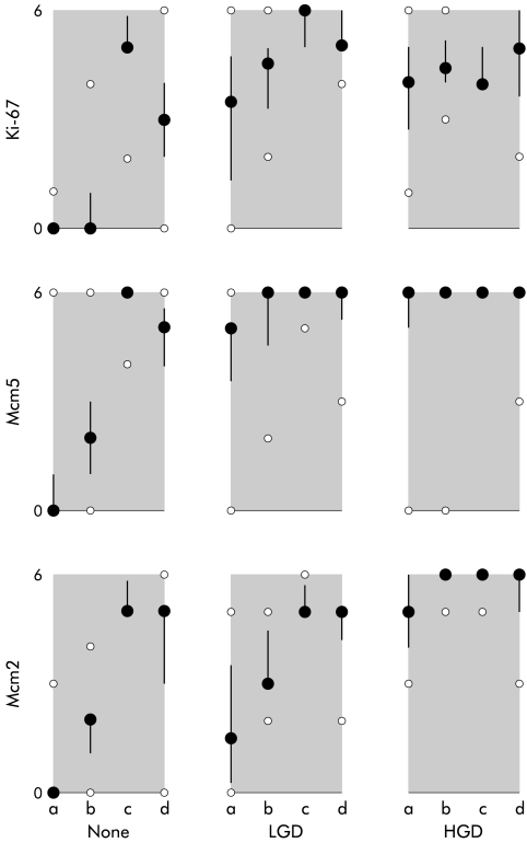
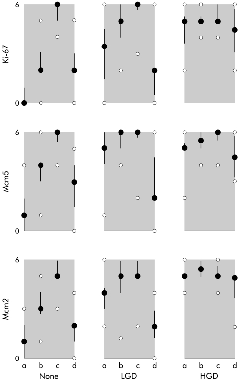
Aim: To test the hypothesis that Mcm proteins are downregulated together with the proliferation marker Ki-67 in differentiating epithelial compartments of non-dysplastic squamous and Barrett's epithelium, and that this process does not occur in dysplastic mucosae. METHODS AND CASES: Forty five patients with Barrett's oesophagus included 20 with glandular dysplasia (10 low grade, eight high grade, two both, and four with invasive adenocarcinoma). Twenty five other patients included 12 with oesophageal squamous dysplasia (three low grade, six high grade, three both, and four with invasive squamous carcinoma). Formalin fixed paraffin embedded tissue sections from biopsy series and resections were immunostained using antibodies to Mcm2, Mcm5, and Ki-67. Percentage of nuclei positive for Mcm2, Mcm5, and Ki-67 was estimated and scored from 0 to 6 as: 0, none +; 1, <10%+; 2, 10-30%+; 3, 30-70%+; 4, 70-90%+; 5, >90%+; 6, all+. Four separate epithelial strata were scored: in squamous epithelium the basal layer and thirds to the surface, in Barrett's mucosa the luminal surface, upper and lower crypt, and deep glands.
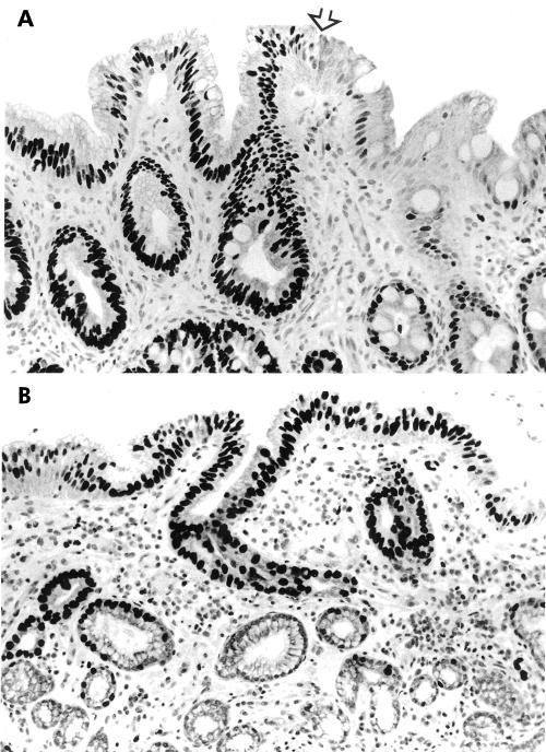
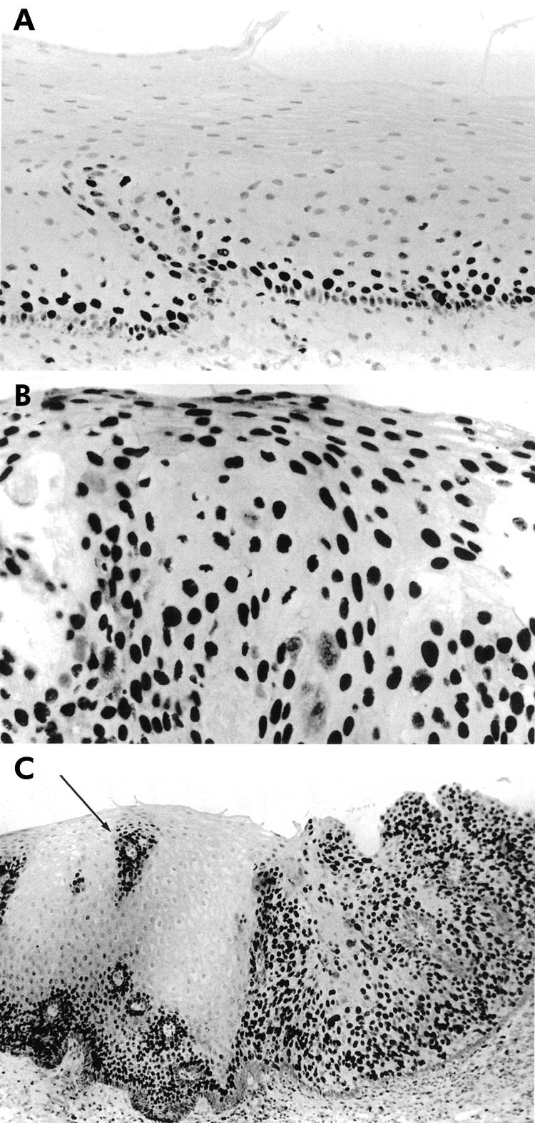
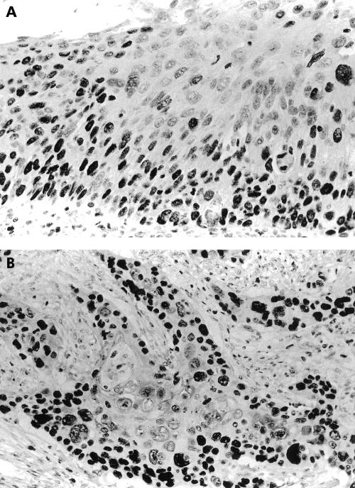
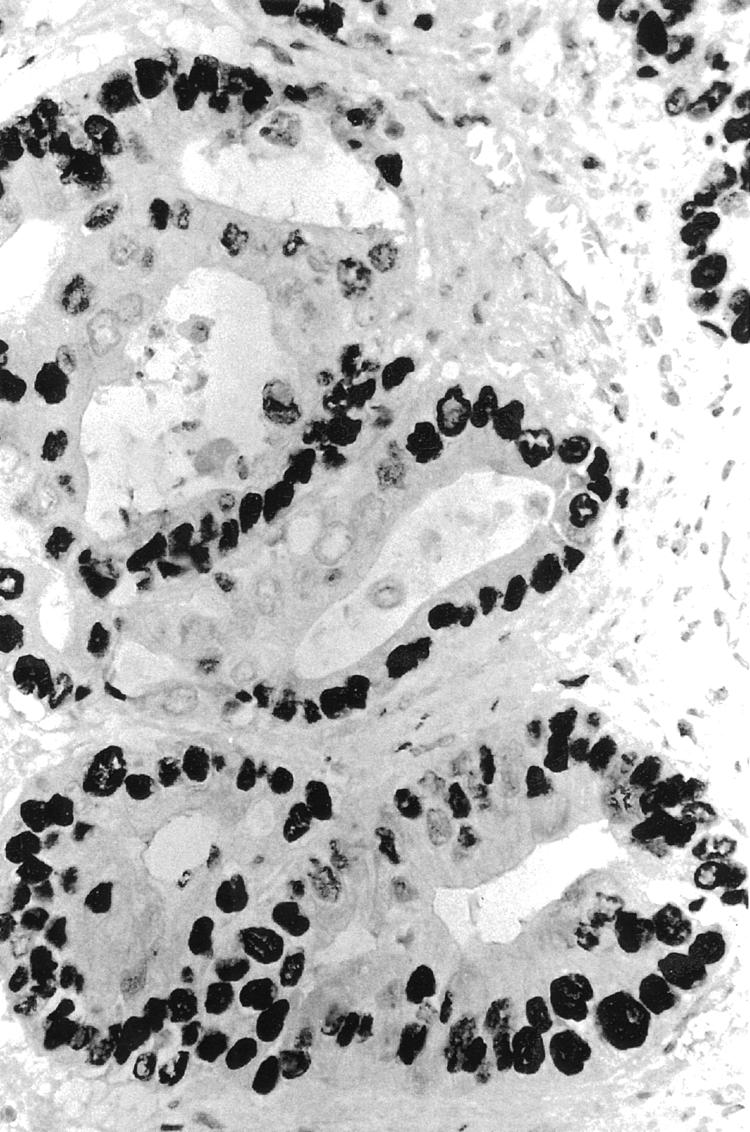
Results: In non-dysplastic squamous epithelium and Barrett's mucosa, high level expression of Mcm2, Mcm5, and Ki-67 proteins was largely confined to the proliferative compartments and downregulated in differentiated compartments. Expression persisted up to the mucosal surface in dysplastic squamous epithelium and Barrett's mucosa.
Conclusions: Persistent expression of Mcm2, Mcm5, and Ki-67 proteins in luminal compartments of dysplastic oesophageal squamous epithelium and dysplastic Barrett's mucosa may be diagnostic markers and imply disruption of cell cycle control and differentiation in these dysplastic epithelia.
Figures
Comment in
-
Minichromosome maintenance (MCM) proteins may be pre-cancer markers.Gut. 2002 Mar;50(3):290-1. doi: 10.1136/gut.50.3.290. Gut. 2002. PMID: 11839701 Free PMC article. No abstract available.
References
-
- Wang HH, Sovie S, Zeroogian JM , et al. Value of cytology in detecting intestinal metaplasia and associated dysplasia at the gastroesophageal junction. Hum Pathol 1997;28:465–71. - PubMed
-
- Yang K, Liu Y, Lipkin M , et al. Precancerous lesions of the human esophagus: multiparametric study of esophageal biopsies from a high-risk population in Linxian, China. J Cell Biochem Suppl 1992;16G:187–94. - PubMed
-
- Shu YJ. Cytopathology of the esophagus. An overview of esophageal cytopathology in China. Acta Cytol 1983;27:7–16. - PubMed
-
- McCarthy M, Wilkinson ML. Treatment of Barrett's esophagus by endoscopic laser ablation and antireflux surgery. Gastrointest Endosc 1999;49:129–30. - PubMed
-
- Dumoulin FL, Terjung B, Neubrand M , et al. Treatment of Barrett's esophagus by endoscopic argon plasma coagulation. Endoscopy 1997;29:751–3. - PubMed
Publication types
MeSH terms
Substances
LinkOut - more resources
Full Text Sources
Other Literature Sources
Medical
Miscellaneous