. 2002 Mar;50(3):392-401.
doi: 10.1136/gut.50.3.392.

Galectin-8 expression decreases in cancer compared with normal and dysplastic human colon tissue and acts significantly on human colon cancer cell migration as a suppressor

Affiliations

Galectin-8 expression decreases in cancer compared with normal and dysplastic human colon tissue and acts significantly on human colon cancer cell migration as a suppressor

N Nagy et al. Gut. 2002 Mar.

Abstract

Background and aims: Galectins are beta-galactoside binding proteins. This ability may have a bearing on cell adhesion and migration/proliferation in human colon cancer cells. In addition to galectins-1 and -3 studied to date, other members of this family not investigated in detail may contribute to modulation of tumour cell features. This evident gap has prompted us to extend galectin analysis beyond the two prototypes. The present study deals with the quantitative determination of immunohistochemical expression of galectin-8 in normal, benign, and malignant human colon tissue samples and in four human colon cancer models (HCT-15, LoVo, CoLo201, and DLD-1) maintained both in vitro as permanent cell lines and in vivo as nude mice xenografts. The role of galectin-8 (and its neutralising antibody) in cell migration was investigated in HCT-15, LoVo, CoLo201, and DLD-1 cell lines.

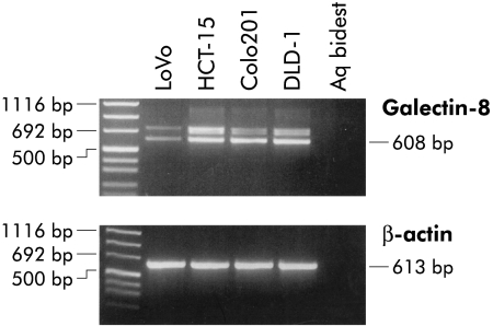
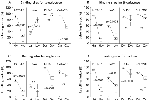
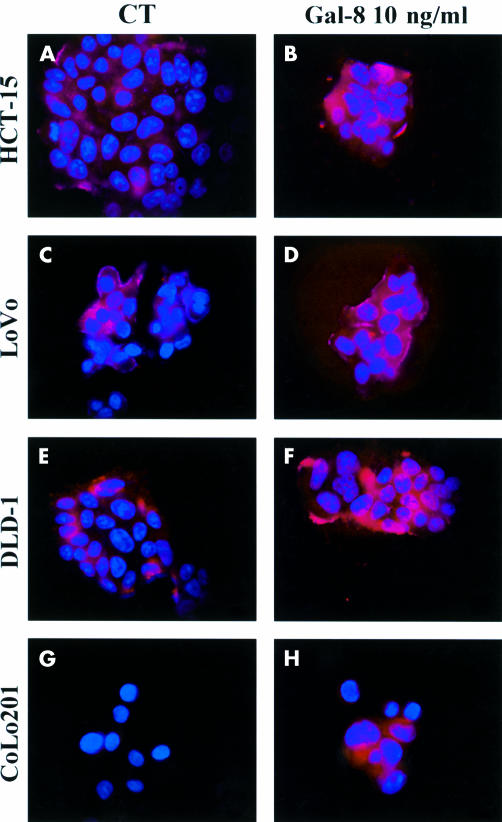
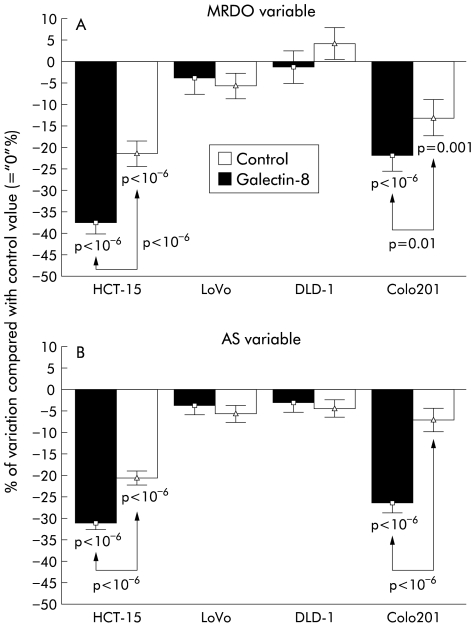
Methods: Immunohistochemical expression of galectin-8 and its overall ability to bind to sugar ligands (revealed glycohistochemically by means of biotinylated histochemically inert carrier bovine serum albumin with alpha- and beta-D-galactose, alpha-D-glucose, and lactose derivatives as ligands) were quantitatively determined using computer assisted microscopy. The presence of galectin-8 mRNA in the four human colon cancer cell lines was examined by reverse transcriptase-polymerase chain reaction. In vitro, cellular localisation of exogenously added galectin-8 in the culture media of these colon cancer cells was visualised by fluorescence microscopy. In vitro galectin-8 mediated effects (and the influence of its neutralising antibody) on migration levels of living HCT-15, LoVo, CoLo201, and DLD-1 cells were quantitatively determined by computer assisted phase contrast microscopy.

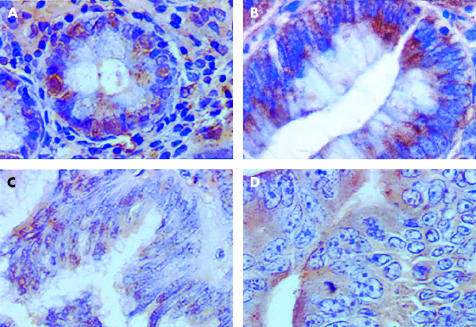
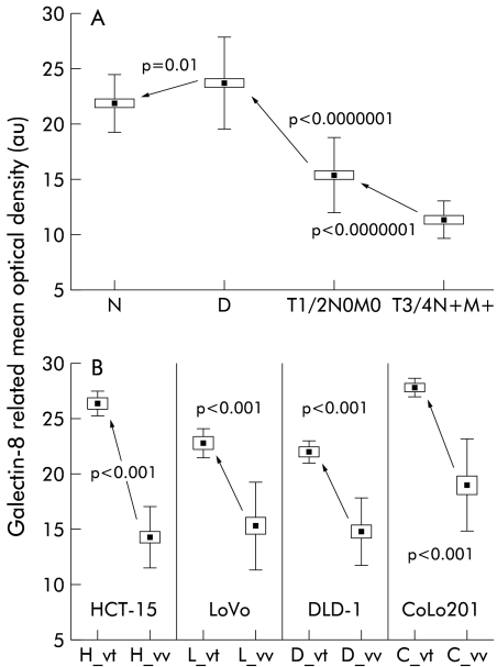
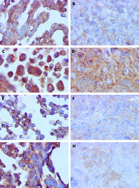
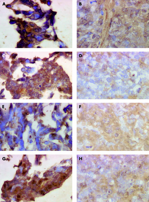
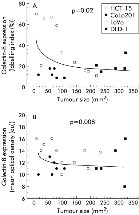
Results: A marked decrease in immunohistochemical expression of galectin-8 occurred with malignancy development in human colon tissue. Malignant colon tissue exhibited a significantly lower galectin-8 level than normal or benign tissue colon cancers; those with extensive invasion capacities (T3-4/N+/M+) harboured significantly less galectin-8 than colon cancers with localised invasion capacities (T1-2/N0/M0). The four experimental models (HCT-15, LoVo, CoLo201, and DLD-1) had more intense galectin-8 dependent staining in vitro than in vivo. Grafting the four experimental human colon cancer models onto nude mice enabled us to show that the immunohistochemical expression of galectin-8 was inversely related to tumour growth rate. In vitro, galectin-8 reduced the migration rate of only those human experimental models (HCT-15 and CoLo201) that exhibited the lowest growth rate in vivo.

Conclusions: Expression of galectin-8 correlated with malignancy development, with suppressor activity, as shown by analysis of clinical samples and xenografts. In vitro, only the two models with low growth rates were sensitive to the inhibitory potential of this galectin. Future investigations in this field should involve fingerprinting of these newly detected galectins, transcending the common focus on galectins-1 and -3.


Figures

Figure 1
Figure 1
Morphological illustration (G×400) of immunohistochemical expression of galectin-8 in glandular tissue from a normal colon (A), adenoma (B), locally invasive carcinoma (C), and an extensively invasive carcinoma (D).
Figure 2
Figure 2
The 41 cases under study were grouped according to different histopathological and clinical variables, and galectin-8 concentration in epithelial tissue (measured as mean optical density) was thus quantitatively determined. (A) Data obtained for the normal (N), dysplastic (D), locally invasive (T1 or T2 and N0 and M0 variables of the TNM staging system), and extensively invasive cancer (T3 or T4 and N+ and M+) cases. (B) Quantitative determination of the extent of galectin-8 expression in epithelial tissue analysed in four human colon cancer cell lines cultured in vitro or grafted subcutaneously into nude mice. Five distinct in vitro cell populations (obtained from five distinct in vitro passages) and five distinct in vivo xenografts were analysed for each of the four models (HCT-15, LoVo, DLD-1, and CoLo201). Ten microscope fields (G×200) were analysed for each tumour. All fields analysed for a given colon model were grouped together. For each of the four models under study, the “x”_vt symbol on the x axis represents the in vitro cell line and the “x”_vv is its corresponding in vivo xenograft. All data are presented as means (black squares), SEM (open rectangles), and SDs (bars).
Figure 3
Figure 3
Galectin-8 gene expression by human colorectal carcinoma cell lines (HCT-15, LoVo, DLD-1, and CoLo201). Reverse transcriptase-polymerase chain reaction analysis revealed the presence of two galectin-8 specific bands in all four cell lines.
Figure 4
Figure 4
Morphological illustration (G×400) of immunohistochemically detected galectin-8 expression in the four human colon cancer cell lines cultured in vitro and in their corresponding in vivo xenografts. (A) In vitro HCT-15 cell line; (B) in vivo HCT-15 xenograft; (C) in vitro CoLo201 cell line; (D) in vivo CoLo201 xenograft; (E) in vitro LoVo cell line; (F) in vivo LoVo xenograft; (G) in vitro DLD-1 cell line; and (H) in vivo DLD-1 xenograft.
Figure 5
Figure 5
Morphological illustration (G×400) of glycohistochemically detected binding sites for four distinct oligosaccharide moieties in the HCT-15 human colon cancer model. (A) (in vitro specimen) and (B) (in vivo specimen) relate to the α-Gal-neoglycoconjugate, (C) (in vitro specimen) and (D) (in vivo specimen) to the β-Gal-neoglycoconjugate, (E) (in vitro specimen) and (F) (in vivo specimen) to the β-Glu-neoglycoconjugate, and (G) (in vitro specimen) and (H) (in vivo specimen) to the Lac-neoglycoconjugate.
Figure 6
Figure 6
The presence of galactose, glucose, and lactose binding sites in the in vitro versus in vivo human HCT-15, LoVo, DLD-1, and CoLo201 colon cancer models was revealed by glycohistochemical probes containing α-galactose (A), β-galactose (B), α-glucose (C), and lactose (D) residues covalently attached as ρ-aminophenyl derivatives to biotinylated otherwise histochemically inert carrier protein. See legend to fig 2B ▶ for abbreviations.
Figure 7
Figure 7
Illustration of galectin-8 binding pattern on human colon cancer cells. Galectin-8 was revealed by fluorescence microscopy with the antigalectin-8 antibody used throughout the study. The four figures on the left (CT panel) illustrate galectin-8 binding patterns in the four cell lines in the culture media to which no exogenous galectin-8 was added. In contrast, the four figures on the right (Gal-8 10 ng/ml panel) correspond to experimental conditions in which 10 ng/ml galectin-8 was added 30 minutes before its visualisation by means of the fluorescent antigalectin-8 antibody.
Figure 8
Figure 8
Data obtained by quantitative video microscopy to compute the maximum relative distance from the point of origin (MRDO variable (A)) and the rate of migration (determined as the average speed (AS) variable (B)) on living HCT-15, LoVo, DLD-1, and CoLo201 human colon cancer cells. The migration levels of these four cell lines were quantitatively assessed in control (arbitrarily defined as “0%” and corresponding to culture supports coated with Matrigel) compared with galectin-8 treated conditions in which galectin-8 was coated onto Matrigel coated culture supports. The potential neutralisation of the biological effects induced by galectin-8 was assayed by adding an antigalectin-8 antibody to the culture media at a concentration of 100 ng/ml. The experiments lasted 48 hours. Data are presented as means (SEM).
Figure 9
Figure 9
Percentage of immunopositive galectin-8 epithelial colon cancer cells (A) and immunohistochemically detected galectin-8 concentration in epithelial colon cancer cells (B) in relation to growth rate in the HCT-15, CoLo201, LoVo, and DLD-1 human colon cancer cell lines grafted into nude mice.

References

    1. Gabius HJ. Endogenous lectins in tumors and the immune system. Cancer Invest 1987;5:39–46. - PubMed
    1. Raz A, Lotan R. Endogenous galactoside-binding lectins: a new class of functional tumor cell surface molecules related to metastasis. Cancer Metastasis Rev 1987;6:433–52. - PubMed
    1. Kim YJ, Varki A. Perspective on the significance of altered glycosylation of glycoproteins in cancer. Glycoconjugate J 1997;14:569–76. - PubMed
    1. Hakomori SI. Cancer-associated glycosphingolipid antigens: their structure, organisation, and function. Acta Anat 1998;161:79–90. - PubMed
    1. Reuter G, Gabius HJ. Eukaryotic glycosylation: whim of nature or multipurpose tool? Cell Mol Life Sci 1999;55:368–22. - PMC - PubMed

Publication types

MeSH terms