. 2002 Feb 18;156(4):751-60.
doi: 10.1083/jcb.200105147. Epub 2002 Feb 11.

Different modes of hypertrophy in skeletal muscle fibers

Affiliations

Different modes of hypertrophy in skeletal muscle fibers

Angelika C Paul et al. J Cell Biol. .

Abstract

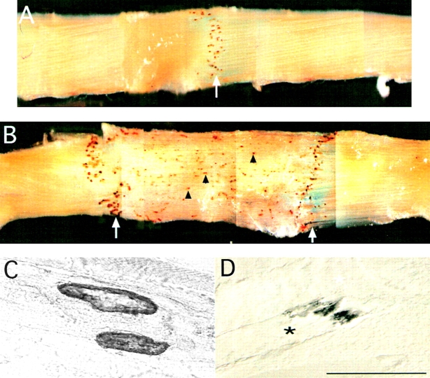
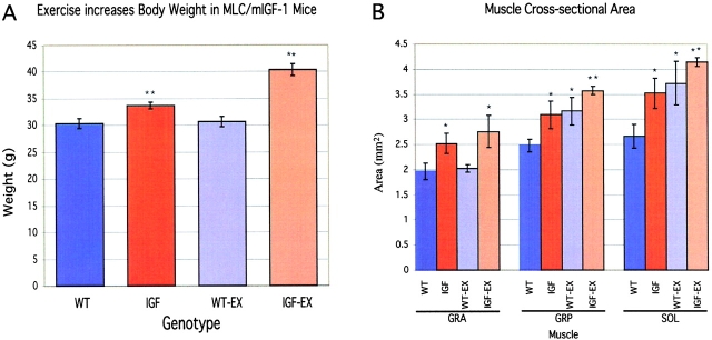
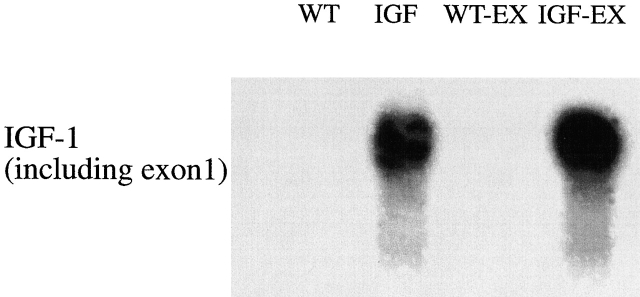
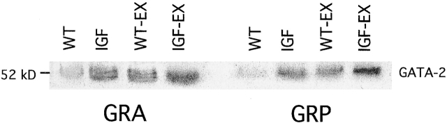
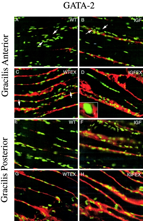
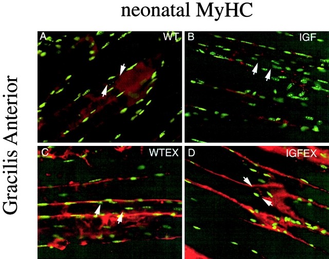
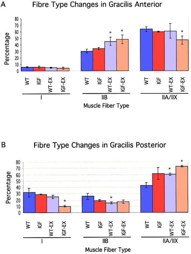
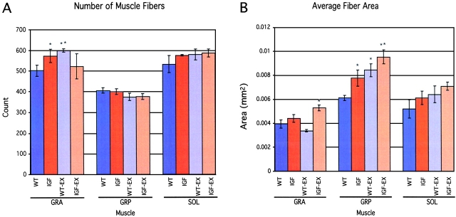
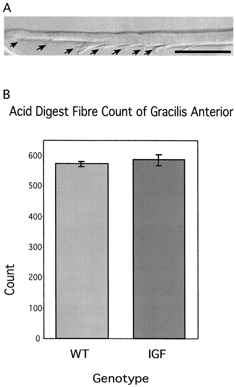
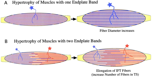
Skeletal muscles display a remarkable diversity in their arrangement of fibers into fascicles and in their patterns of innervation, depending on functional requirements and species differences. Most human muscle fascicles, despite their great length, consist of fibers that extend continuously from one tendon to the other with a single nerve endplate band. Other mammalian muscles have multiple endplate bands and fibers that do not insert into both tendons but terminate intrafascicularly. We investigated whether these alternate structural features may dictate different modes of cell hypertrophy in two mouse gracilis muscles, in response to expression of a muscle-specific insulin-like growth factor (IGF)-1 transgene (mIGF-1) or to chronic exercise. Both hypertrophic stimuli independently activated GATA-2 expression and increased muscle cross-sectional area in both muscle types, with additive effects in exercising myosin light chain/mIGF transgenic mice, but without increasing fiber number. In singly innervated gracilis posterior muscle, hypertrophy was characterized by a greater average diameter of individual fibers, and centralized nuclei. In contrast, hypertrophic gracilis anterior muscle, which is multiply innervated, contained longer muscle fibers, with no increase in average diameter, or in centralized nuclei. Different modes of muscle hypertrophy in domestic and laboratory animals have important implications for building appropriate models of human neuromuscular disease.


Figures

Figure 1.
Figure 1.
AChE precipitate showing pattern of innervation in singly and multiply innervated muscles. (A) Gracilis posterior muscles of the mouse have a single endplate band in the center of the muscle (arrow) and no intrafascicularly terminating fibers. (B) Gracilis anterior muscle of the mouse has two endplate bands (arrows) and intrafascicularly terminating fibers whose ends also show the AChE precipitate (arrowheads). (C) Neuromuscular junction labeled with AChE precipitate. (D) Myo–myonal junction between two intrafascicularly terminating fibers (white and black asterisks) labeled with AChE precipitate.
Figure 2.
Figure 2.
Skeletal muscle hypertrophy of inactive and exercised WT and mIGF transgenic mice. (A) Comparison of body weights shows a significant increase in IGF and IGF-EX compared with WT animals, with IGF-EX animals weighing the most. (B) Muscle cross-sectional area increased significantly in gracilis anterior, gracilis posterior, and soleus muscles, indicating hypertrophy in these muscles. Since the MLC/mIGF-1 transgene is expressed at very low levels in soleus (Musarò et al., 2001) the increase in the average cross-sectional area of soleus muscle may reflect a secondary effect of increased loading in neighboring muscles, resulting in a transition to a faster fiber type.
Figure 3.
Figure 3.
Northern blot showing levels of mIGF-1 in thigh muscles from WT, IGF, WT-EX, and IGF-EX using a the full-length probe against the tissue-restricted isoform of IGF-1. This week-long exposure shows no IGF-1 RNA in WT and WT-EX muscles. Only in the MLC/mIGF-1 transgenic mice is the gene expressed, with an upregulation in IGF-EX. The endogenous gene is not expressed in any specimen. The endogenous IGF-1 gene is therefore not upregulated with voluntary wheel-running exercise. The transgene is under the control of the MLC promoter and is upregulated with exercise.
Figure 4.
Figure 4.
Western blot using an antibody against GATA-2 transcription factor, a marker for skeletal muscle hypertrophy. In both gracilis anterior and posterior the WT muscle contained the least amount of GATA-2 protein, followed by an equal level in IGF and WT-EX muscles. IGF-EX muscles had the highest expression of GATA-2 protein, indicating that exercise and the mIGF-1 transgene synergize to increase muscle hypertrophy.
Figure 5.
Figure 5.
GATA-2 localization on gracilis anterior or posterior muscles with Hoechst (green fluorescence) and AChE (brightfield) stains. (A–D) Gracilis Anterior with AChE precipitate between arrows. (A) WT, (B) MLC/mIGF-1 transgenic, (C) WT-EX, (D) MLC/mIGF-1 transgenic exercised, and (D, insert) GATA-2 is excluded from the nucleus. (E–H) Gracilis posterior. (E) WT, (F) MLC/mIGF-1 transgenic, (G) WT-EX, (H) MLC/mIGF-1 transgenic exercised. GATA-2 protein is upregulated in IGF and WT-EX, with a further increase in IGF-EX.
Figure 6.
Figure 6.
Neonatal myosin localization on gracilis anterior muscles with Hoechst (green fluorescence) and AChE (brightfield) stains. (A–D) Gracilis anterior with AChE precipitate between arrows. (A) WT, (B) MLC/mIGF-1 transgenic, (C) WT-EX, (D) MLC/mIGF-1 transgenic exercised. Neonatal myosin is upregulated along the length of fibers in WT-EX and IGF-EX muscles.
Figure 7.
Figure 7.
Trend in fiber type changes in gracilis anterior and posterior muscle influenced by exercise not transgene. (A) Gracilis anterior muscle showed a trend of increasing type IIB MyHC at the cost of type IIA/X MyHC–positive fibers. A significant difference was only found in WT-EX and transgenic mice. (B) Fiber type changes in gracilis posterior muscle showed a trend toward type IIA/X MyHC–positive fibers at the cost of fibers expressing type IIB and type I MyHC. Exercise appears to be the main influence on these trends.
Figure 8.
Figure 8.
Number and diameter of muscle fibers. (A) The number of muscle fibers increased significantly only in gracilis anterior muscle and not gracilis posterior or soleus muscles. (B) The average fiber area increased significantly in gracilis posterior muscle and in IGF-EX gracilis anterior muscle only. Singly innervated muscles therefore respond to hypertrophic signals by increasing average fiber area and not number. Gracilis anterior muscle, which contains intrafascicularly terminating fibers, responds to hypertrophic signals by increasing the number of muscle fibers in cross-section.
Figure 9.
Figure 9.
Counts of total fiber number in gracilis anterior after acid digestion. (A) Acid digestion of gracilis anterior muscle separates intrafascicularly terminating fibers that taper in steps (arrows). (B) Counts of all muscles fibers in WT and mIGF-1 transgenic mice after acid digest does not show a difference in total fiber number. Therefore, the increase in fiber number seen in cross-section is the result of elongation of existing intrafascicularly terminating muscle fibers.
Figure 10.
Figure 10.
Model of modes of skeletal muscle hypertrophy. (A) Skeletal muscles with a single endplate band hypertrophy by increasing the diameter of individual fibers. (B) Intrafascicularly terminating fibers (IFTs) in a muscle with two endplate bands hypertrophy by elongating, causing an increase in the number of fibers in transverse section (TS).
Figure 11.
Figure 11.
Percentage of muscle fibers with centralized nuclei in gracilis anterior and posterior muscles. In the gracilis posterior the number of centralized nuclei was increased in mIGF animals, with a large increase in the IGF-EX muscle, indicating hypertrophy along the length of fibers. In contrast, the number of centralized nuclei was not increased in gracilis anterior muscle.

References

    1. Adams, D., and B. MacKay. 1961. The distribution of motor end-plates in mammalian muscles. Bibl. Anat. 2:153–154.
    1. Allen, D.L., B.C. Harrison, A. Maass, M.L. Bell, W.C. Byrnes, and L.A. Leinwand. 2001. Cardiac and skeletal muscle adaptations to voluntary wheel running in the mouse. J. Appl. Physiol. 90:1900–1908. - PubMed
    1. Barton-Davis, E.R., D.I. Shoturma, A. Musaro, N. Rosenthal, and H.L. Sweeney. 1998. Viral mediated expression of insulin-like growth factor I blocks the aging-related loss of skeletal muscle function. Proc. Natl. Acad. Sci. USA. 95:15603–15607. - PMC - PubMed
    1. Brown, I.E., T. Satoda, F.J. Richmond, and G.E. Loeb. 1998. Feline caudofemoralis muscle. Muscle fibre properties, architecture, and motor innervation. Exp. Brain Res. 121:76–91. - PubMed
    1. Clark, G. 1981. Methods for connective tissue. Staining Procedures. G. Clark, editor. Williams & Wilkins, Baltimore. 512.

Publication types

MeSH terms