. 2002 Feb 18;156(4):609-15.
doi: 10.1083/jcb.200110068. Epub 2002 Feb 11.

Force transduction by Triton cytoskeletons

Affiliations

Force transduction by Triton cytoskeletons

Yasuhiro Sawada et al. J Cell Biol. .

Abstract

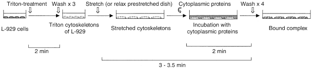
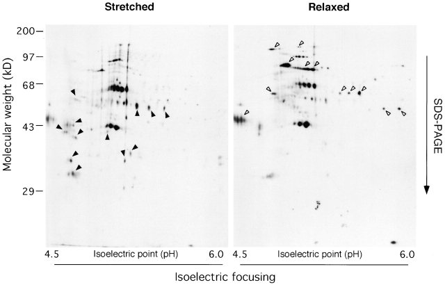
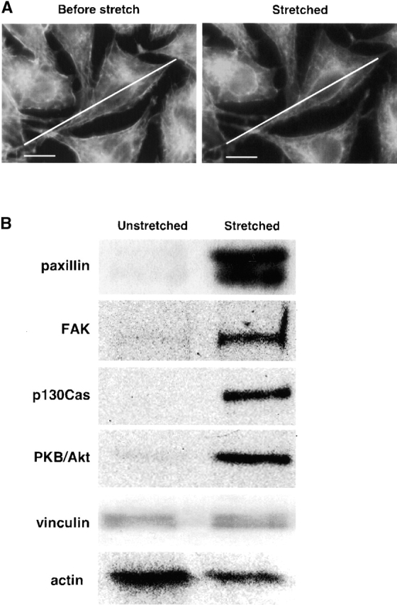
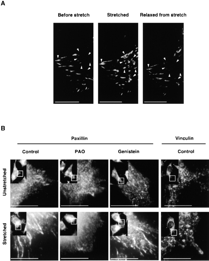
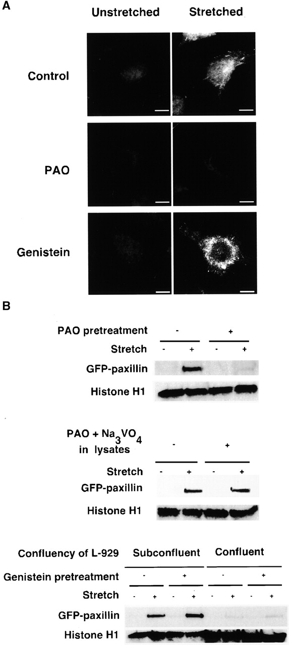
Force-initiated signal transduction can occur either via membrane-based ionic mechanisms or through changes in cytoskeletal-matrix linkages. We report here the stretch-dependent binding of cytoplasmic proteins to Triton X-100 cytoskeletons of L-929 cells grown on collagen-coated silicone. Triton X-100-insoluble cytoskeletons were stretched by 10% and incubated with biotinylated cytoplasmic proteins. Analysis with two-dimensional gel electrophoresis showed stretch-dependent binding of more than 10 cytoplasmic protein spots. Bound cytoplasmic proteins were purified by a photocleavable biotin tag and stretch-dependent binding of paxillin, focal adhesion kinase, and p130Cas was found, whereas the binding of vinculin was unchanged and actin binding decreased with stretch. Paxillin binding upon stretch was morphologically and biochemically similar in vitro and in vivo, that is, enhanced in the periphery and inhibited by the tyrosine phosphatase inhibitor, phenylarsine oxide. Thus, we suggest that transduction of matrix forces occurs through force-dependent conformation changes in the integrated cytoskeleton.


Figures

Figure 1.
Figure 1.
Diagram of protocol for stretch-dependent binding of cytoplasmic proteins to Triton X-100–insoluble cytoskeletons. L-929 cells were cultured on a collagen-coated silicone substrate, and cytoskeletons were prepared by treating with 0.25% Triton X-100/ISO (+) buffer for 2 min (as described in Materials and methods). Triton X-100–insoluble cytoskeletons were either left unstretched or stretched (or relaxed from prestretch) with ISO (+) after washing three times. Then, the ISO (+) buffer was replaced with the cytoplasmic lysate solution (as described in Materials and methods), incubated for 2 min at room temperature, and washed four times with ISO (+) buffer.
Figure 2.
Figure 2.
2-D gels of biotinylated proteins that bound to stretched or relaxed cytoskeletons. The complex of the cytoskeleton with the biotinylated cytoplasmic proteins (Fig. 1) was solubilized with 1 ml of a rehydration buffer (8 M urea, 2% CHAPS, 20 mM DTT, 0.5% IPG buffer [Amersham Pharmacia Biotech]) for isoelectrical focusing (the first dimension of 2-D gel electrophoresis). Immobiline dry strip (pH 4–7; Amersham Pharmacia Biotech) was rehydrated with 350 μl of each sample and subjected to isoelectrical focusing followed by SDS-PAGE. Biotinylated cytoplasmic proteins in 2-D gels were visualized with affinity blotting using HRP-conjugated Streptavidin. Arrowheads mark the spots that were found specifically in Stretched or Relaxed.
Figure 3.
Figure 3.
Focal contact proteins bind preferentially to stretched cytoskeletons. (A) Micrographs showing that Triton X-100–insoluble cytoskeletons are stretched 10%. Triton X-100–insoluble cytoskeletons on a collagen-coated silicone membrane (StageFlexer®; Flexcell International) were incubated with rhodamine-phalloidin (Molecular Probes) for 2 min, and washed three times with ISO (+) buffer. Images were obtained with an Olympus BX50 microscope with a 60×, 0.9 NA water immersion objective. Focus was adjusted to identify the peripheral margins (lower surface of cells), and images were obtained before stretch (Before stretch) and 5 min after stretch (Stretched). The diagonal lines show the length of the cell before stretch. (B) Western blots of focal contact proteins bound to unstretched and stretched cytoskeletons. L-929 cytoplasmic proteins tagged with a photocleavable biotin (NHS-PC-LC-biotin) were added to Triton X-100–insoluble cytoskeletons of L-929 cells on a stretchable silicone dish (Sawada et al., 2001), and cytoskeletons were stretched or left unstretched (Fig. 1). After washing, bound cytoplasmic proteins were eluted with 1 ml of 1 M NaCl in HYPO buffer (as described in Materials and methods), precipitated with avidin beads (immobilized neutravidin; Pierce Chemical Co.) after sevenfold dilution with HYPO buffer, and released from the bead complex by irradiation with 302 nm UV light (10 min). After photocleavage, proteins were eluted with 120 μl of HYPO buffer, and 40 μl of the sample was subjected to 10% SDS-PAGE followed by immunoblotting with antibodies to paxillin, FAK, p130Cas, PKB/Akt (Transduction Laboratories), vinculin (Upstate Biotechnology), or actin (Santa Cruz Biotechnology). Bar, 10 μm.
Figure 4.
Figure 4.
Fluorescence micrographs of the stretch-dependent distribution of GFP paxillin, endogenous paxillin, and vinculin in intact L-929 cells. (A) L-929 cells transiently transfected with GFP paxillin were cultured on collagen-coated silicone membranes in a StageFlexer system. Cells were stretched by 10%, held for 5 min, and subsequently relaxed by allowing the stretched silicone membrane to return to its original size. GFP fluorescence was observed with an Olympus BX50 microscope using a 60×, 0.9 NA water immersion objective before stretch (left), 2 min after stretch (middle), and 2 min after relaxation of stretch (right). Arrowheads indicate sites of GFP paxillin assembly dynamics: Before stretch, Stretched, and Relaxed from stretch. (B) Antipaxillin or antivinculin antibody distribution in L-929 cells cultured on silicone membranes was measured either for unstretched (top), stretched (for 2 min; bottom), stretched PAO treated (20 μM, for 10 min), or stretched genistein treated (100 μM, for 10 min) cells. Cells were fixed with 3.7% formaldehyde/PBS, permeabilized with 0.1% Triton X-100/PBS, and subjected to immunostaining using an antipaxillin or an antivinculin antibody. Stretched samples were relaxed after fixation. The rectangle in the inset (low magnification) indicates the area of each micrograph. Bars, 10 μm.
Figure 5.
Figure 5.
In vitro GFP paxillin binding to Triton X-100–insoluble cytoskeletons. (A) Triton X-100–insoluble cytoskeletons of L-929 cells in a StageFlexer system either unstretched (left) or stretched (10%; right) were incubated with 293-GFP-pax lysates (supplemented with 0.5 mM ATP, 2% BSA) for 2 min. After four washes with ISO (+) buffer, the bound complex of cytoplasmic proteins and cytoskeletons (Fig. 1) was fixed with 3.7% formaldehyde/ISO buffer. Stretched samples were relaxed after fixation, and the distribution of bound GFP paxillin was visualized with fluorescence microscopy (Olympus BX50 microscope with a 60×, 0.9 NA water immersion objective). PAO (20 μM; center) or genistein (100 μM; bottom) was added 10 min before permeabilization and stretching. (B) Triton X-100–insoluble cytoskeletons of L-929 cells in silicone dishes either unstretched or stretched by 10% were incubated with 293-GFP-pax lysates for 2 min. After four washes with ISO (+) buffer, the bound complex of cytoplasmic proteins and cytoskeletons (Fig. 1) were solubilized with 500 μl of SDS sample buffer (100 mM Tris-HCl, pH 6.8, 36% glycerol, 4% SDS, 10 mM DTT, 0.01% bromophenol blue). 50 μl of each sample was subjected to SDS-PAGE followed by immunoblotting with antibodies to GFP (CLONTECH Laboratories, Inc.) and histone H1 (Santa Cruz Biotechnology, Inc.). PAO (20 μM; top), genistein (100 μM; bottom), or solvent (0.1% DMSO) was added 10 min before permeabilization and stretching. Triton X-100–insoluble cytoskeletons were prepared from 4 × 10⁵ cells per dish with the exception of the confluent culture (four right lanes on bottom) where 1.2 × 10⁶ cells per dish were used. Bar, 10 μm.

References

    1. Akiyama, T., J. Ishida, S. Nakagawa, H. Ogawara, S. Watanabe, N. Itoh, M. Shibuya, and Y. Fukami. 1987. Genistein, a specific inhibitor of tyrosine-specific protein kinases. J. Biol. Chem. 262:5592–5595. - PubMed
    1. Avnur, Z., J.V. Small, and B. Geiger. 1983. Actin-independent association of vinculin with the cytoplasmic aspect of the plasma membrane in cell-contact areas. J. Cell Biol. 96:1622–1630. - PMC - PubMed
    1. Balaban, N.Q., U.S. Schwarz, D. Riveline, P. Goichberg, G. Tzur, I. Sabanay, D. Mahalu, S. Safran, A. Bershadsky, L. Addadi, and B. Geiger. 2001. Force and focal adhesion assembly: a close relationship studied using elastic micropatterned substrates. Nat. Cell Biol. 3:466–472. - PubMed
    1. Ball, E.H., C. Freitag, and S. Gurofsky. 1986. Vinculin interaction with permeabilized cells: disruption and reconstitution of a binding site. J. Cell Biol. 103:641–648. - PMC - PubMed
    1. Bang, O.S., E.J. Kim, J.G. Chung, S.R. Lee, T.K. Park, and S.S. Kang. 2000. Association of focal adhesion kinase with fibronectin and paxillin is required for precartilage condensation of chick mesenchymal cells. Biochem. Biophys. Res. Commun. 278:522–529. - PubMed

Publication types

MeSH terms

Substances