. 2002 Mar;22(5):1329-39.
doi: 10.1128/MCB.22.5.1329-1339.2002.

Yeast protein kinases and the RHO1 exchange factor TUS1 are novel components of the cell integrity pathway in yeast

Affiliations

Yeast protein kinases and the RHO1 exchange factor TUS1 are novel components of the cell integrity pathway in yeast

Tobias Schmelzle et al. Mol Cell Biol. 2002 Mar.

Abstract

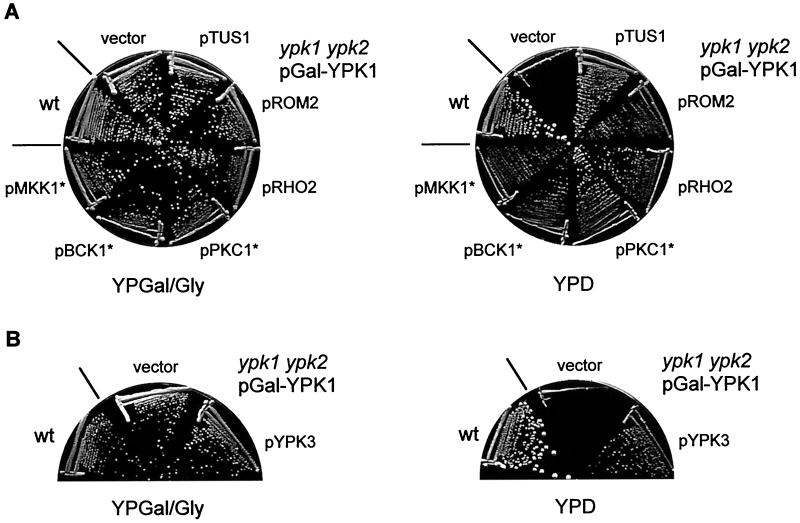
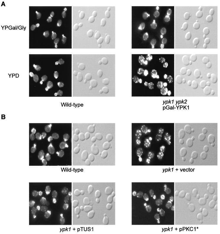
The PKC1-associated mitogen-activated protein (MAP) kinase pathway of Saccharomyces cerevisiae regulates cell integrity by controlling the actin cytoskeleton and cell wall synthesis. Activation of PKC1 occurs via the GTPase RHO1 and the kinase pair PKH1 and PKH2. Here we report that YPK1 and YPK2, an essential pair of homologous kinases and proposed downstream effectors of PKH and sphingolipids, are also regulators of the PKC1-controlled MAP kinase cascade. ypk mutants display random distribution of the actin cytoskeleton and severely reduced activation of the MAP kinase MPK1. Upregulation of the RHO1 GTPase switch or the PKC1 effector MAP kinase pathway suppresses the growth and actin defects of ypk cells. ypk lethality is also suppressed by overexpression of an uncharacterized gene termed TUS1. TUS1 is a novel RHO1 exchange factor that contributes to cell wall integrity-mediated modulation of RHO1 activity. Thus, TUS1 and the YPKs add to the growing complexity of RHO1 and PKC1 regulation in the cell integrity signaling pathway. Furthermore, our findings suggest that the YPKs are a missing link between sphingolipid signaling and the cell integrity pathway.


Figures

FIG. 1.
FIG. 1.
Activation of the RHO1 GTPase switch or the PKC1 effector MAP kinase cascade suppresses the growth defect of ypk cells. (A) Wild-type (wt) (TB50a) cells and ypk1 ypk2/pGal-YPK1 (TS57-2B) cells transformed with either empty vector, pTUS1, pROM2, pRHO2, pPKC1*, pBCK1*, or pMKK1* were streaked onto YPGal/Gly (galactose) and YPD (glucose) media and incubated at 37°C. (B) Overexpression of YPK3 restores growth of ypk cells. wt (TB50a) cells and ypk1 ypk2/pGal-YPK1 (TS57-2B) cells transformed with empty vector or pYPK3 were streaked onto YPGal/Gly and YPD media and incubated at 30°C.
FIG. 2.
FIG. 2.
Genetic interaction of TUS1 with components of the cell integrity pathway. (A) The tus1/tus1 mutant is defective in cell integrity signaling. Wild-type (wt) (JK9-3da/α) cells and tus1/tus1 (TS49) cells transformed with either empty vector, pROM2, pRHO2, or pPKC1* were streaked out onto YPD medium and incubated at 30 and 39°C. (B) Osmotic stabilization suppresses the growth defect of the tus1/tus1 mutant. wt (JK9-3da/α) and tus1/tus1 (TS49) cells were streaked out onto YPD medium and YPD medium containing 1 M sorbitol at 39°C. (C) wt (TB50a), tus1 (TS24-6D), rom2 (SD40-1A), tus1 rom2 (TS63-2B), rho2 (TS40-5B), tus1 rho2 (TS43-2B), bck1 (PA109-1C), tus1 bck1 (TS100-1B), mpk1 (TS45-1A), and tus1 mpk1 (TS48-3A) cells were streaked out onto YPD medium and incubated at 37°C. Strains were also streaked out onto YPD medium containing 1 M sorbitol at 37°C.
FIG. 3.
FIG. 3.
TUS1 is an exchange factor for RHO1. (A) TUS1 interacts with nucleotide-free RHO1 in the two-hybrid system. Yeast strain EGY191 was transformed with pEG202::TUS1ΔN and pJG4-5 (vector), pJG4-5::RHO1, pJG4-5::RHO1Q68H, or pJG4-5::RHO1G22A and streaked out onto SGal/Raff-Ura-Trp-His/X-Gal plates at 30°C. Blue color (seen here as dark color) indicates a specific interaction in the two-hybrid system. (B) The TUS1 DH domain mediates the TUS1-RHO1 interaction. Yeast strain EGY191 was transformed with pJG4-5::RHO1G22A and pEG202 (vector), pEG202::TUS1ΔN, or TUS1 fragments in pEG202 (pTS49, pTS50, and pTS56) and incubated on SGal/Raff-Ura-Trp-His/X-Gal plates at 30°C. A schematic representation of the TUS1 bait constructs is shown on the right; numbers represent amino acid positions. Blue color (seen here as dark color) indicates a specific interaction in the two-hybrid system. (C) TUS1 has GEF activity toward RHO1. GST-RHO1 was preincubated with GDP and subsequently mixed with [35S]GTPγS and GST-TUS1DH (circles) or GST (squares). Reactions were stopped after 5, 10, and 15 min of incubation, and reaction products were washed. [35S]GTPγS bound to GST-RHO1 was quantified by scintillation counting (see Materials and Methods). A representative curve obtained from one of at least three experiments is shown.
FIG. 4.
FIG. 4.
Activation of RHO1 in response to cell wall defects requires TUS1. (A) Wild-type (wt) (JK9-3da) cells, tor2ts (SH121) cells, and tus1 tor2ts (TS27-4C) cells were streaked onto YPD medium and onto YPD medium containing 0.005% SDS and were incubated at 37°C. As a control, strains were also streaked onto YPD medium containing 0.005% SDS and incubated at 30°C. (B) wt (JK9-3da), tus1 cwh41 (TS80-6D), tor2ts (SH121), cwh41 tor2ts (MB119), and tus1 cwh41 tor2ts (TS92-2D) cells were streaked onto YPD medium and incubated at 30 and 37°C. A tus1 mutation prevents the suppression of the tor2 mutation by cell wall defects (induced by SDS or cwh41).
FIG. 5.
FIG. 5.
ypk cells display an actin organization defect. (A) Wild-type (TB50a) and ypk1 ypk2/pGal-YPK1 (TS57-2B) cells were grown in YPGal/Gly (galactose) medium, shifted to YPD (glucose) medium for 16 h, fixed, stained for actin with TRITC-phalloidin, and observed by fluorescence (actin, left panels) and Nomarski (right panels) microscopy. (B) Overexpression of TUS1 or expression of PKC1* suppresses the actin defect in ypk1 cells. Wild-type (TB50a) cells and ypk1 (TS38-1C) cells transformed with empty vector, pTUS1, or pPKC1* were pregrown in SD medium. Cells were then grown to early-logarithmic phase in YPD medium and processed for actin staining. Fluorescence (actin) and Nomarski microscopy images are shown in the left and right panels, respectively.
FIG. 6.
FIG. 6.
YPK1 is required for MPK1 activation in response to heat stress. Wild-type (TS99-5C) and ypk1 (TS105-1C) cells, expressing MPK1-HA3, were grown to early-logarithmic phase at 24°C (time 0), shifted to 39°C for the indicated times (20, 40, 60, 80, 100, and 120 min), and processed for total protein extraction, SDS-PAGE, and Western analysis as described in Materials and Methods. The upper panel for each strain is the anti-activated MAP kinase immunoblot (phospho-MPK1); the lower panel is the anti-HA immunoblot (MPK1-HA3).

References

    1. Alberts, A. S., N. Bouquin, L. H. Johnston, and R. Treisman. 1998. Analysis of RhoA-binding proteins reveals an interaction domain conserved in heterotrimeric G protein beta subunits and the yeast response regulator protein Skn7. J. Biol. Chem. 273:8616-8622. - PubMed
    1. Alberts, A. S., and R. Treisman. 1998. Activation of RhoA and SAPK/JNK signalling pathways by the RhoA-specific exchange factor mNET1. EMBO J. 17:4075-4085. - PMC - PubMed
    1. Ausubel, F. M., R. Brent, R. E. Kingston, D. D. Moore, J. G. Seidman, J. A. Smith, and K. Struhl. 1998. Current protocols in molecular biology. John Wiley & Sons, Inc., New York, N.Y.
    1. Beck, T., A. Schmidt, and M. N. Hall. 1999. Starvation induces vacuolar targeting and degradation of the tryptophan permease in yeast. J. Cell Biol. 146:1227-1238. - PMC - PubMed
    1. Bell, L. M., M. L. Leong, B. Kim, E. Wang, J. Park, B. A. Hemmings, and G. L. Firestone. 2000. Hyperosmotic stress stimulates promoter activity and regulates cellular utilization of the serum- and glucocorticoid-inducible protein kinase (Sgk) by a p38 MAPK-dependent pathway. J. Biol. Chem. 275:25262-25272. - PubMed

Publication types

MeSH terms

Substances