. 2002 Mar;22(5):1495-503.
doi: 10.1128/MCB.22.5.1495-1503.2002.

Conditional loss of Nkx3.1 in adult mice induces prostatic intraepithelial neoplasia

Affiliations

Conditional loss of Nkx3.1 in adult mice induces prostatic intraepithelial neoplasia

Sarki A Abdulkadir et al. Mol Cell Biol. 2002 Mar.

Abstract

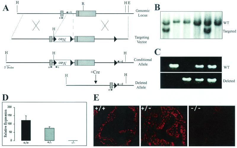
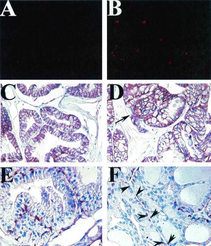
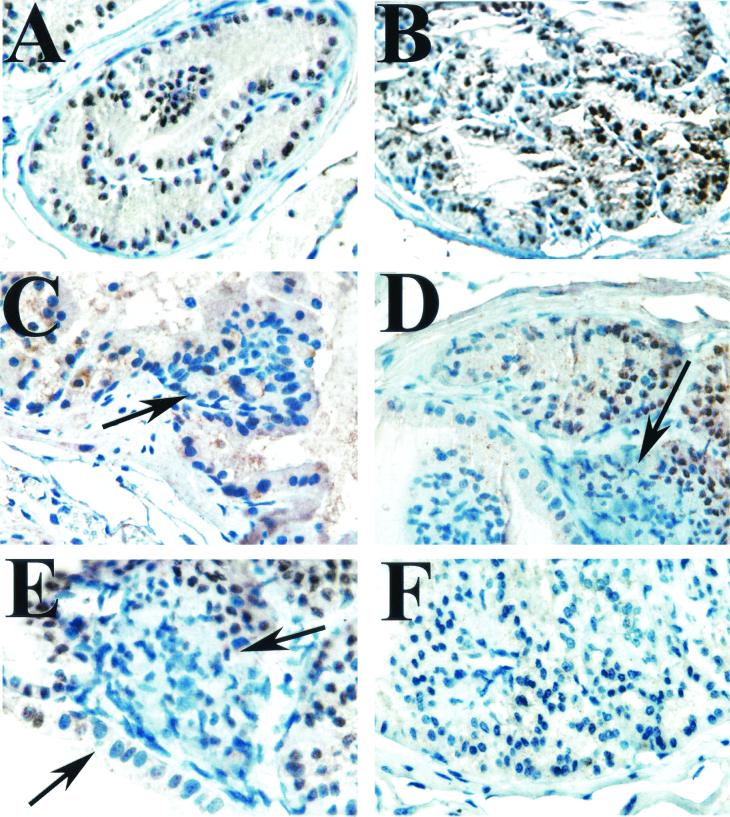
The homeodomain-containing transcription factor NKX3.1 is a putative prostate tumor suppressor that is expressed in a largely prostate-specific and androgen-regulated manner. Loss of NKX3.1 protein expression is common in human prostate carcinomas and prostatic intraepithelial neoplasia (PIN) lesions and correlates with tumor progression. Disruption of the murine Nkx3.1 gene results in defects in prostate branching morphogenesis, secretions, and growth. To more closely mimic the pattern of NKX3.1 loss that occurs in human prostate tumors, we have used Cre- and loxP-mediated recombination to delete the Nkx3.1 gene in the prostates of adult transgenic mice. Conditional deletion of one or both alleles of Nkx3.1 leads to the development of preinvasive lesions that resemble PIN. The pattern of expression of several biomarkers (Ki-67, E-cadherin, and high-molecular-weight cytokeratins) in these PIN lesions resembled that observed in human cases of PIN. Furthermore, PIN foci in mice with conditional deletion of a single Nkx3.1 allele lose expression of the wild-type allele. Our results support the role of NKX3.1 as a prostate tumor suppressor and indicate a role for this gene in tumor initiation.


Figures

FIG. 1.
FIG. 1.
Generation of conditional and knockout Nkx3.1 alleles. (A) Maps of the Nkx3.1 genomic locus and the loxP-targeting vector designed to introduce a PGK-NeoR cassette flanked by two loxP sites and a third loxP site into the 3" UTR. Exons are denoted by rectangles and loxP sites are denoted by black triangles. The cross-hatched rectangles indicate coding regions, while the solid rectangle denotes the 3" UTR. Successful targeting yields a conditional (Nkx3.1flox) allele. Cre-mediated recombination results in deletion of the PGK-neor cassette and the homeodomain-containing second exon, yielding a knockout or deleted allele. Specific primers used to screen for the various alleles are indicated by small arrows labeled a through d. H, HindIII; R, EcoRI; E, EcoRI. (B) Southern blot analysis performed with a 5" external probe to detect homologous recombination resulting in the generation of a conditional (Nkx3.1flox) allele. This probe detects a 9-kb HindIII wild-type (WT) fragment and a 6-kb fragment from the targeted allele. (C) PCR analysis of genomic DNA from wild-type (Nkx3.1+/+), heterozygous (Nkx3.1+/−), and knockout (Nkx3.1−/−) mice generated by first mating mice containing the conditional allele (Nkx3.1flox) with Actin-Cre transgenic deleter mice (21) and subsequent mating of the heterozygous F1 animals. (D) Quantitative RT-PCR analysis of Nkx3.1 expression in mouse prostates with the indicated genotypes. (E) Immunohistochemical analysis of Nkx3.1 expression in the anterior prostates of wild-type, heterozygous, and knockout mice. Note that the heterozygous prostate is hyperplastic, explaining the multiple layers of Nkx3.1-positive luminal epithelial nuclei. Original magnifications, ×400.
FIG. 2.
FIG. 2.
Analysis of the prostates of conventional Nkx3.1-deficient mice. (A to F) Hematoxylin- and eosin-stained sections of anterior prostates from 20-week-old Nkx3.1+/+(A and D), Nkx3.1+/− (B and E), and Nkx3.1−/− mice (C and F). Note the hyperplasia evident in sections from Nkx3.1+/− and Nkx3.1−/− mice. (G to I) Analysis of proliferation by Ki-67 staining of Nkx3.1+/+(G), Nkx3.1+/− (H), and Nkx3.1−/− (I) prostates. Magnification for panels A to C, ×100. Magnification for panels D to I, ×400.
FIG. 3.
FIG. 3.
Generation and characterization of PSA-Cre transgenic mice. (A and B) Cell-type-specific expression of Nkx3.1 protein in the adult prostate. (A) NKX3.1 protein expression is restricted to the luminal epithelial cells of human prostate glands (brown nuclei) but not basal cells or stromal cells (blue nuclei). (B) Immunohistochemical localization of Nkx3.1 expression to luminal epithelial cell nuclei in mouse prostate (red). Basal cells stained with antibody to high-molecular-weight cytokeratins, 34βE12 (green), are negative for Nkx3.1 expression. (C to E) Assay of Cre activity in PSA-Cre;Z/AP double transgenic mice by double lacZ/alkaline phosphatase staining. The prostate of a control 8-week-old Z/AP-positive, PSA-Cre-negative transgenic mouse (C) shows widespread lacZ activity (blue) in the epithelium. The sections in panels D and E show the prostate of an 8-week-old double transgenic PSA-Cre;Z/AP animal demonstrating replacement of lacZ staining with alkaline phosphatase expression (pink) in luminal epithelial cells in which Cre-mediated recombination has occurred. (F) Quantitative RT-PCR (TaqMan) analysis for expression of the Cre transgene in prostates of mice from the two PSA-Cre transgenic lines (Cre3 and Cre13) and a normal prostate (N).
FIG. 4.
FIG. 4.
Temporal and prostate lobe-specific pattern of Cre activity in PSA-Cre transgenic mice. (A) Age-dependent Cre activity in PSA-Cre+ Nkx3.1flox transgenic mice. Cre-mediated recombination, detected by PCR analysis of genomic DNA, is evident in the prostates of 10-week- and 30-week-old mice but not in the prostates of 3-week-old animals. (B) Detection of Cre-mediated recombination in various tissues of a 30-week-old PSA-Cre+ Nkx3.1flox/flox transgenic mouse by PCR. R, recombined allele; UR, unrecombined allele; Cre, PSA-Cre transgene; AP, anterior prostate; DLP, dorsolateral prostate; VP, ventral prostate; SV, seminal vesicle; T, testis; B, brain; K, kidney; L, liver; H, heart; w, water control.
FIG. 5.
FIG. 5.
Prostatic intraepithelial neoplasia and epithelial hyperplasia in conditional Nkx3.1-deficient mice. (A) Prostate section from a control 28-week-old Nkx3.1+/flox mouse showing normal epithelium. (B to F) Epithelial hyperplasia and PIN (arrows) in the prostates of a 28-week-old PSA-Cre+ Nkx3.1+/flox mouse (B), a 23-week-old PSA-Cre+ Nkx3.1flox/− mouse (C), a 15-week-old PSA-Cre+ Nkx3.1+/flox mouse (D and E), and 25-week-old PSA-Cre+ Nkx3.1flox/flox transgenic mice (F through I). Original magnification for panels A to C, ×400; for panels D, F, G, and H, ×200; for panels E and I, ×600.
FIG. 6.
FIG. 6.
Characterization of PIN lesions that develop in conditional Nkx3.1-deficient mice. (A and B) Ki-67 immunohistochemistry, showing increased labeling in a PIN lesion from the prostate of a PSA-Cre+ Nkx3.1flox/flox mouse (B) relative to that in a normal prostate (A). (C and D) Pattern of E-cadherin expression by immunohistochemistry in normal prostatic epithelium (C) and a PIN lesion from a PSA-Cre+ Nkx3.1flox/flox mouse (D). Note the retention of membrane E-cadherin staining in the PIN lesion. The arrow in panel D points to a rare example of early microinvasion. (E and F) Staining of basal cells with antibody against high-molecular-weight cytokeratins (34βE12) in normal prostate (E) compared to that in conditional Nkx3.1-deficient PIN lesions (F). Note partial disruption of the basal cell layer in PIN lesions (arrows in panels F).
FIG. 7.
FIG. 7.
Immunohistochemical analysis of Nkx3.1 expression in conditional Nkx3.1-deficient mice. (A) Nkx3.1 expression in a benign prostate gland showing nuclear staining (brown) of the luminal epithelial cells. Hematoxylin was used as the counterstain (blue). (B) Hyperplastic glands from a PSA-Cre+ Nkx3.1+/flox mouse showing that these cells have largely retained expression of Nkx3.1 (brown nuclei). (C to E) PIN lesions from PSA-Cre+ Nkx3.1+/flox mice showing cells that have lost expression of Nkx3.1 (blue nuclei, arrows) consistent with loss or silencing of the wild-type allele. (F) A lesion from a PSA-Cre+ Nkx3.1flox/flox transgenic mouse showing complete loss of Nkx3.1 expression.

References

    1. Abdulkadir, S. A., G. F. Carvalhal, Z. Kaleem, W. Kisiel, P. A. Humphrey, W. J. Catalona, and J. Milbrandt. 2000. Tissue factor expression and angiogenesis in human prostate carcinoma. Hum. Pathol. 31:443-447. - PubMed
    1. Abdulkadir, S. A., Z. Qu, E. Garabedian, S. K. Song, T. J. Peters, J. Svaren, J. M. Carbone, C. K. Naughton, W. J. Catalona, J. J. Ackerman, J. I. Gordon, P. A. Humphrey, and J. Milbrandt. 2001. Impaired prostate tumorigenesis in Egr1-deficient mice. Nat. Med. 7:101-107. - PubMed
    1. Bhatia-Gaur, R., A. A. Donjacour, P. J. Sciavolino, M. Kim, N. Desai, P. Young, C. R. Norton, T. Gridley, R. D. Cardiff, G. R. Cunha, C. Abate-Shen, and M. M. Shen. 1999. Roles for Nkx3.1 in prostate development and cancer. Genes Dev. 13:966-977. - PMC - PubMed
    1. Bieberich, C. J., K. Fujita, W. W. He, and G. Jay. 1996. Prostate-specific and androgen-dependent expression of a novel homeobox gene. J. Biol. Chem. 271:31779-31782. - PubMed
    1. Bostwick, D. G., D. Ramnani, and J. Qian. 2000. Prostatic intraepithelial neoplasia: animal models 2000. Prostate 43:286-294. - PubMed

Publication types