. 2002 Mar;22(5):1504-12.
doi: 10.1128/MCB.22.5.1504-1512.2002.

Mouse Pop1 is required for muscle regeneration in adult skeletal muscle

Affiliations

Mouse Pop1 is required for muscle regeneration in adult skeletal muscle

Birgit Andrée et al. Mol Cell Biol. 2002 Mar.

Abstract

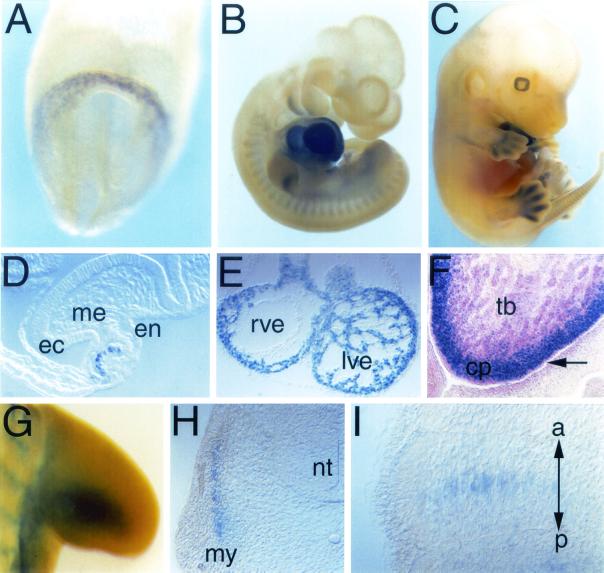
Popeye (Pop) genes are a novel gene family encoding putative transmembrane proteins predominantly present in striated and smooth muscle cells. In this study, a null mutation of Pop1 was generated by replacing the first coding exon of the Pop1 gene with the lacZ reporter gene. Homozygous mice lacking Pop1 were fertile and had a normal life span without any apparent phenotype. LacZ staining of tissues of heterozygous and homozygous Pop1-LacZ mice revealed strong expression in embryonic and fetal hearts. Pop1-LacZ was also expressed in the myotome and in myogenic progenitor cells within the limb and in smooth muscle cells of various organs. In the heart, Pop1-LacZ activity was downregulated postnatally in heterozygous mice but not in homozygous mice. Administration of the beta-adrenergic agonist isoproterenol led to a rapid increase in Pop1-LacZ activity in heterozygotes without induction at the transcriptional level, suggesting stabilization of the protein. No difference, however, was observed between homozygous and heterozygous mice in the ability to develop cardiac hypertrophy in response to isoproterenol. The capacity to regenerate skeletal muscle was tested after cardiotoxin injection into the hind limbs of hetero- and homozygous mice. In activated satellite cells of both genotypes, rapid activation of Pop1-LacZ expression was observed. In heterozygous animals, LacZ activity was only transiently elevated in muscle precursor cells undergoing fusion and in newly formed myotubes. In homozygotes, persistence of LacZ expression and a retarded ability to regenerate skeletal muscle were apparent, suggesting that Pop1 plays a role in muscle regeneration.


Figures

FIG. 1.
FIG. 1.
Targeted disruption of Pop1. (A) Restriction map of the Pop1 locus and gene-targeting strategy. The targeting vector was constructed by replacing the coding region of the first coding exon with an NLS-lacZ cassette (β-galactosidase-encoding gene with a simian virus 40 NLS at the 5" end). 3" of the NLS-lacZ gene was a neomycin resistance-encoding gene (neo) in the opposite orientation with respect to the coding region of the Pop1 gene. The left homology arm was a 6.2-kb EcoRI genomic fragment. For the right homology arm, a 1-kb genomic fragment was amplified by PCR. Homologous recombinants were identified by Southern blot analysis using a 200-bp fragment (So) amplified by PCR from genomic DNA (hybridizing to the Pop1 gene 3" to the targeted region). Hybridization of DNA digested with XbaI with So generated 5- and 8-kb fragments from the mutant and wild-type alleles, respectively. Exons are represented by filled boxes. E, EcoRI; X, XbaI; hsv-tk, herpes simplex virus thymidine kinase. (B) Offspring were genotyped by Southern blot analysis. Lanes 1, 4, and 8 contained DNAs from homozygotes, lanes 2, 3, 5, and 6 contained samples from heterozygotes, and lane 7 contained DNA from a wild-type animal. (C) RT-PCR analysis of adult heart, skeletal muscle (skm), and bladder reveals an absence of the Pop1 transcript in mutant mice. Pop3 expression appeared not to be altered in cardiac and skeletal muscle in the absence of Pop1; however, Pop3 was slightly upregulated in the homozygote bladder.
FIG. 2.
FIG. 2.
Pop1-LacZ expression in heterozygous Pop1-LacZ mouse embryos. LacZ expression was detected after whole-mount staining (A to C and G) or after cryosectioning (F). (D and E) Cryosections of the cardiac region of the embryos shown in panels A and B, respectively. (A and D) E7.5 embryo. LacZ activity is detectable in the cardiogenic mesoderm of the anterior (presumptive ventricular) heart field. (B) E10.5 embryo. Expression is detectable in the heart, branchial arches, and somites and in a posterior domain within the limb. (C) E13.5 embryo. Expression is found in the heart and the peridigital mesenchyme. (E) Section through the heart of the embryo shown in panel B. Myocytes were LacZ positive, while endocardial and epicardial cells were devoid of Pop1-LacZ staining. (F) Section through the heart of an E13.5 embryo stained for Pop1-LacZ activity and counterstained with eosin. Pop1-LacZ activity was confined to the compact layer (cp) myocardium and was strongly diminished or absent from myocytes in the trabecular layer (tb). (G) E10.5 embryo. Pop1-LacZ activity was present within the limb in a posterior domain. (H and I) Transverse (H) and sagittal (I) sections through somites of a E10.5 embryo. Staining is confined to the myotome. The two-headed arrow in panel I indicates the anterior and posterior borders of the somite. Pop1-LacZ appears to label forming myotubes in the center of the somite. ec, ectoderm; en, endoderm; me, mesoderm; my, myotome; nt, neural tube; rve, right ventricle; lve, left ventricle.
FIG. 3.
FIG. 3.
Pop1-LacZ expression in postnatal hearts from heterozygous Pop1-LacZ mice. LacZ expression was detected in frozen sections of postnatal day 1 (A and A"), 3 (B and B"), and 8 (C and C") and adult (3 months old; D and D") hearts. Panels A" to D" are high-power views of sections of hearts of the same ages as those shown in panels A to D that were stained for β-galactosidase activity. The section in panel A" was incubated for 5 h, while the sections shown in panels B" to D" were incubated for 16 h in order to visualize staining in myocyte nuclei (arrows) and cytoplasmic vesicles (arrowheads), respectively. (E) RT-PCR analysis of Pop1, Pop1-LacZ, and glyceraldehyde-3-phosphate dehydrogenase (GAPDH) expression in cardiac muscle at postnatal days 1, 8, 14, 90, and 240.
FIG. 4.
FIG. 4.
Pop1-LacZ activity in heart and skeletal muscles of heterozygous (A, C, and E) and homozygous (B, D, and F) Pop1-LacZ animals. Panels: A and B, ventricle; C and D, atrium; E and F, skeletal muscle.
FIG. 5.
FIG. 5.
Pop1-LacZ activity in ventricular tissue of heterozygous Pop1-LacZ animals after 3 days of infusion of PBS (A) and Iso (B). (C) RT-PCR analysis of Pop1-LacZ. Pop1, GATA4, ANF, and RPL7 mRNA expression in ventricular tissue of PBS- and Iso-infused animals 3 days after pump implantation. The arrows in panel B indicate nuclear LacZ activity.
FIG. 6.
FIG. 6.
Morphological and molecular analysis of skeletal muscle after injection of cardiotoxin. (A) Pop1-LacZ activity in heterozygous (+/−) and homozygous (−/−) mice at 5, 10, and 20 days after cardiotoxin injection. (B) RT-PCR analysis of myogenin, MyoD, Myf5, MRF4, Pop1, Pop1-LacZ, and glyceraldehyde-3-phosphate dehydrogenase (GAPDH) in skeletal muscle tissue that was excised at the indicated times after cardiotoxin injection.
FIG. 7.
FIG. 7.
Differentiation of adult satellite cells isolated from skeletal muscle of heterozygous (+/−) and homozygous (−/−) Pop1-LacZ mice. (A and E) Nuclear Pop1-LacZ activity in satellite cells cultured in growth medium. (B and F) Expression of M-cadherin (brown staining) and Pop1-LacZ in satellite cells cultured in growth medium. Arrowheads demarcate cells that express M-cadherin but not Pop1-LacZ. (C and G) Pop1-LacZ expression in cells cultured for 3 days in differentiation medium. In satellite cells derived from heterozygous mice (C), LacZ activity is downregulated in myotubular nuclei (arrowhead) while myoblasts display nuclear LacZ (arrows). In contrast, in satellite cells derived from homozygous muscle (G), nuclear LacZ activity is present in both unfused myoblasts (arrow in the middle) and myotubes (arrows on the left and right). (D and H) Satellite cells cultured for 5 days in differentiation medium and stained for MF20 and Pop1-LacZ. Irrespective of the genotype, nuclear LacZ staining is lost from myotubes (arrowheads); however, myoblasts (arrows) display nuclear lacZ expression.

References

    1. Allen, R., C. Temm-Grove, S. Sheehan, and G. Rice. 1997. Methods in muscle biology. Methods Cell Biol. 52:155-176. - PubMed
    1. Andrée, B., T. Hillemann, G. Kessler-Icekson, T. Schmitt-John, H. Jockusch, H. H. Arnold, and T. Brand. 2000. Isolation and characterization of the novel popeye gene family expressed in skeletal muscle and heart. Dev. Biol. 223:371-382. - PubMed
    1. Brand, T., H. S. Sharma, and W. Schaper. 1993. Expression of nuclear proto-oncogenes in isoproterenol-induced cardiac hypertrophy. J. Mol. Cell. Cardiol. 25:1325-1337. - PubMed
    1. d'Albis, A., R. Couteaux, C. Janmot, and J. C. Mira. 1989. Myosin isoform transitions in regeneration of fast and slow muscles during postnatal development of the rat. Dev. Biol. 135:320-325. - PubMed
    1. Danto, S. I., and D. A. Fischman. 1984. Immunocytochemical analysis of intermediate filaments in embryonic heart cells with monoclonal antibodies to desmin. J. Cell Biol. 98:2179-2191. - PMC - PubMed

Publication types

MeSH terms