. 2002 Feb 15;30(4):958-65.
doi: 10.1093/nar/30.4.958.

The MT domain of the proto-oncoprotein MLL binds to CpG-containing DNA and discriminates against methylation

Affiliations

The MT domain of the proto-oncoprotein MLL binds to CpG-containing DNA and discriminates against methylation

Marco Birke et al. Nucleic Acids Res. .

Abstract

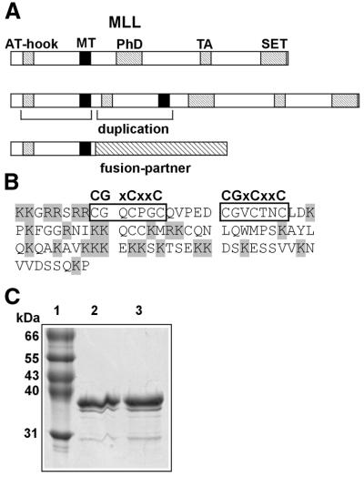
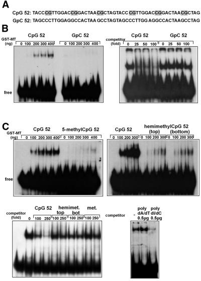
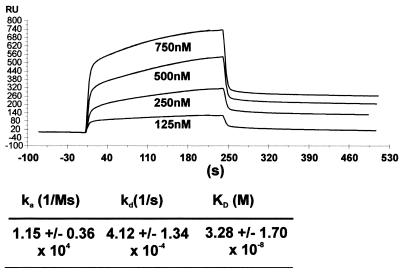
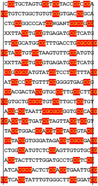
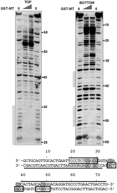
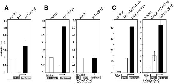
Alterations of the proto-oncogene MLL (mixed lineage leukemia) are characteristic for a high proportion of acute leukemias, especially those occurring in infants. The activation of MLL is achieved either by an internal tandem duplication of 5' MLL exons or by chromosomal translocations that create chimeric proteins with the N-terminus of MLL fused to a variety of different partner proteins. A domain of MLL with significant homology to the eukaryotic DNA methyltransferases (MT domain) has been found to be essential for the transforming potential of the oncogenic MLL derivatives. Here we demonstrate that this domain specifically recognizes DNA with unmethylated CpG sequences. In gel mobility shifts, the presence of CpG was sufficient for binding of recombinant GST-MT protein to DNA. The introduction of 5-methylCpG on one or both DNA strands precluded an efficient interaction. In surface plasmon resonance a KD of approximately 3.3 x 10(-8) M was determined for the GST-MT/DNA complex formation. Site selection experiments and DNase I footprinting confirmed CpG as the target of the MT domain. Finally, this interaction was corroborated in vivo in reporter assays utilizing the DNA-binding properties of the MT domain in a hybrid MT-VP16 transactivator construct.


Figures

Figure 1
Figure 1
Structure of MLL, the MT domain and purification of GST–MT protein. (A) Graphical representation (not to scale) of MLL and its oncogenic derivatives. Conserved domains are indicated as hatched bars, the MT domain is depicted in black. The oncogenic alterations are indicated. AT-hook, AT-hook DNA-binding motifs; PhD, plant homeodomain protein–protein interaction motif; TA, transacting domain interacting with CBP; SET, conserved SET domain associates with chromatin remodeling complexes. (B) Amino acid sequence of the MT domain as used in the experiments as a fusion with GST. The CGxCxxC core is boxed, basic amino acids are shaded. (C) Coomassie-stained SDS–polyacrylamide gel showing purified GST–MT fusion protein. The sizes of the molecular weight standards are indicated to the left. Lane 1, standard; lanes 2 and 3, elution fractions (5 µg protein each) after affinity chromatography.
Figure 2
Figure 2
GST–MT protein binds to unmethylated CpG sequences in DNA. (A) Sequence of the probes used in gel mobility shift analysis. CpG sequences are shaded. (B) Mobility shift experiment (left) and competition experiment (right). For the mobility shift the indicated amounts of GST–MT protein were allowed to bind either to the CpG52 probe or the CpG-free GpC52 probe. Complexes formed were separated in a polyacrylamide gel and visualized by autoradiography. In the competition experiment 400 ng of GST–MT were reacted with labeled CpG52 probe in the presence of the indicated molar excess of unlabeled competitor (either CpG52 or GpC52). (C) Mobility shift experiment with methylated DNA (top). The complex formation of GST–MT protein and methylated DNA was investigated as described for (B). To this purpose either an unmodified probe (CpG52) or a probe with identical sequence but 5mC replacing the cytosine of CpG dinucleotides was used. The label 5-methylCpG52 indicates that the probe was methylated on both strands; hemimethylCpG52 signifies a probe methylated in the top or bottom strand only (as indicated). Competition experiments with methylated DNA (bottom). The complex formation of 400 ng of GST–MT with labeled, unmethylated CpG52 probe was challenged with the indicated excess of unlabeled oligos (left) or homopolymers of DNA (right). The competitor DNA used was either parental CpG52 oligo, CpG52 hemimethylated in the top or bottom strand, completely methylated CpG52 oligo, or homopolymeric DNA consisting of poly-desoxyinosine/desoxycytidine or poly-desoxyadenine/desoxythymidine.
Figure 3
Figure 3
Kd determination for the GST–MT/DNA interaction by BIAcore experiments. The CpG52 oligonucleotide probe was immobilized on a plasmon resonance sensor chip and the kinetic association/dissociation constants were determined in a flow cell charged with the given concentrations of GST–MT protein. The curves show the graphical representation of the measurements in a typical experiment. The kinetic values represent the mean ± SD (n = 8).
Figure 4
Figure 4
GST–MT prefers CpG sequences in site selection experiments. Site selection experiments (Materials and Methods) were performed with oligonucleotides containing a core of 26 random nt. The sequences of 19 clones with high affinity to GST–MT are shown. The vertical lines indicate the extent of the original randomly designed sequence with flanking nucleotides given if they contribute to form a CpG dinucleotide. CpG sequences are boxed.
Figure 5
Figure 5
GST–MT protects CpG from DNase I digestion. The DNase I footprinting pattern of GST–MT bound to clone 1 (as identified in the site selection experiment) was determined. A corresponding DNA fragment was labeled with [γ-33P]ATP either at the top or the bottom strand, complexed with increasing amounts of GST–MT and subjected to DNase I digestion. The reaction products were separated on a denaturing polyacrylamide gel and visualized by autoradiography. The numbers indicate the respective position of the nucleotides and the vertical bars indicate regions protected by GST–MT. The results are overlaid onto the sequence of the probe in a graphical representation. CpGs in the probe are shaded, protected areas are indicated by boxes and hypersensitive areas are marked by a line on the appropriate DNA strand. The open box at the top strand from nucleotides 20 to 30 signifies that the 5′ extent of the footprint could not be exactly determined due to the proximity of the labeled DNA end to the protected area.
Figure 6
Figure 6
A fusion of the MT domain with the VP16 transactivator associates with DNA inside living cells. (A) An expression construct coding for MT and a MT–VP16 fusion was cotransfected into 293T cells with a reporter construct containing a luciferase gene under the control of the SV40 minimal promoter (graphical representation under the respective diagram). As control, empty vector was used as indicated. Luciferase activities were determined 48 h after transfection, normalized to protein content and the respective values are given as relative units with the basal SV40 driven luciferase levels set to one unit. The means ± SD of three independent experiments are given. (B) Expression plasmids as in (A) were cotransfected with a reporter containing an increased CpG content (SV40 promoter preceded by six repetitions of the CpG-rich sequence of site selection clone 1). Either unmodified plasmid (left) or DNA pre-methylated at CpG sequences with SssI methylase was used. (C) Control constructs encoding a fusion of the GAL4 DNA-binding domain and either the MT–VP16 chimera or the VP16 transactivator were transfected as in (A). An unmethylated (left) or pre-methylated (right) plasmid with the SV40 promoter preceded by three copies of the GAL4 DNA-binding consensus site served as a reporter.

References

    1. Dimartino J.F. and Cleary,M.L. (1999) Mll rearrangements in haematological malignancies: lessons from clinical and biological studies. Br. J. Haematol., 106, 614–626. - PubMed
    1. Look A.T. (1997) Oncogenic transcription factors in the human acute leukemias. Science, 278, 1059–1064. - PubMed
    1. Tkachuk D.C., Kohler,S. and Cleary,M.L. (1992) Involvement of a homolog of Drosophila trithorax by 11q23 chromosomal translocations in acute leukemias. Cell, 71, 691–700. - PubMed
    1. Pirrotta V. (1997) Chromatin-silencing mechanisms in Drosophila maintain patterns of gene expression. Trends Genet., 13, 314–318. - PubMed
    1. Pirrotta V. (1998) Polycombing the genome: PcG, trxG and chromatin silencing. Cell, 93, 333–336. - PubMed

Publication types

MeSH terms