. 2002 Feb 19;99(4):1915-20.
doi: 10.1073/pnas.022664799. Epub 2002 Feb 12.

Soybean ENOD40 encodes two peptides that bind to sucrose synthase

Affiliations

Soybean ENOD40 encodes two peptides that bind to sucrose synthase

Horst Rohrig et al. Proc Natl Acad Sci U S A. .

Abstract

ENOD40 is expressed at an early stage in root nodule organogenesis in legumes. Identification of ENOD40 homologs in nonleguminous plants suggests that this gene may have a more general biological function. In vitro translation of soybean ENOD40 mRNA in wheat germ extracts revealed that the conserved nucleotide sequence at the 5' end (region I) encodes two peptides of 12 and 24 aa residues (peptides A and B). These peptides are synthesized de novo from very short, overlapping ORFs. Appropriate ORFs are present in all legume ENOD40s studied thus far. In this case small peptides are directly translated from polycistronic eukaryotic mRNA. The 24-aa peptide B was detected in nodules by Western blotting. Both peptides specifically bind to the same 93-kDa protein, which was affinity purified from soybean nodules. Using peptide mass fingerprinting, we identified this binding protein as nodulin 100, which is a subunit of sucrose synthase. Based on our data we suggest that ENOD40 peptides are involved in the control of sucrose use in nitrogen-fixing nodules.


Figures
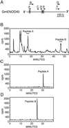
Figure 1
Figure 1
HPLC analyses of the in vitro translation products synthesized from GmENOD40 mRNA. (A) Location of possible ORFs and conserved regions in GmENOD40 mRNA. Short ORFs are indicated by arrows. The conserved regions I and II are boxed. (B) Strong cation-exchange chromatography of the extracted in vitro translation products on a polysulfoethyl aspartamide column. SCX chromatography was carried out with a 60-min linear salt gradient at a flow rate of 0.7 ml/min. Radiolabeled peptides A and B were collected as indicated. (C and D) Zorbax C-18 RP-HPLC of the radioactive fractions indicated in B. The column was eluted with a linear acetonitrile gradient at a flow rate of 0.8 ml/min. The chromatograms in B, C, and D show radioactivity detected by an on-line radioactivity monitor.
Figure 2
Figure 2
Immunoprecipitation of in vitro translation products. [35S]-Methionine-labeled peptides translated in wheat germ extracts from wild-type GmENOD40 mRNA were solid-phase extracted (lane 1, control), immunoprecipitated with antigen-specific IgGs (lanes 2 and 3), and analyzed by Tricine-SDS/16.5% PAGE and autoradiography. For immunoprecipitation, affinity-purified Abs directed against ORF A- and ORF B-deduced synthetic peptides were used.
Figure 3
Figure 3
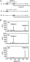
Translation of mutant mRNAs in wheat germ cell-free system in the presence of anti-peptide Abs. (A) Schematic presentation of two small overlapping ORFs of region I located at the 5′ end of wild-type (wt) GmENOD40 mRNA (ORF A and ORF B, Fig. 1A). Mutations in the initiation codons of ORF B and ORF A are indicated (mutant mRNAs M1 and M2). (B) In vitro translation of wt and mutant mRNAs in the presence of antipeptide A and antipeptide B Abs (50 μg each) and SCX chromatography of radiolabeled products.
Figure 4
Figure 4
Detection of peptide B in nodules. Samples were analyzed by tricine-SDS/16.5% PAGE, transferred to a poly(vinylidene difluoride) membrane, and detected with peptide B-specific Abs. Arrowhead marks the position of peptide B.
Figure 5
Figure 5
Peptide A and peptide B bind to the same target protein in nodule extracts. Analysis by SDS/PAGE. Biotinylated peptides A and B (lanes 1 and 3) and the corresponding biotinylated control peptides (lanes 2 and 4) were incubated with extracts from soybean nodules and subsequently bound to streptavidin-agarose beads. Proteins isolated with the peptide affinity matrix were eluted, analyzed by SDS/10% PAGE, and stained with SYPRO ruby. The arrowhead marks the position of the 93-kDa peptide-binding protein. Sizes of molecular mass markers (lane M) are indicated.
Figure 6
Figure 6
Binding of wild-type and mutant GmENOD40 peptides A and B to SuSy. (A) SuSy was partially purified from soybean nodule extract by gel chromatography on Superdex 200. The SuSy-containing fraction used for binding reactions (bar) and the elution position of marker proteins ferritin, catalase, lactate dehydrogenase, and lysozyme are indicated (arrowheads). (B and C) Binding of biotinylated peptides and mutant derivatives to SuSy. Substitutions of amino acid residues in the corresponding peptides are underlined. SuSy was isolated from the Superdex fraction (lane 1) with peptide-streptavidin-agarose beads. Proteins bound to the affinity matrix were eluted by boiling in sample buffer, resolved by SDS/10% PAGE, and stained with SYPRO ruby. Lane M, molecular size markers. The authenticity of the SuSy protein band was verified by Western blotting with anti-SuSy Abs (data not shown).

References

    1. Minami E, Kouchi H, Cohn J R, Ogawa T, Stacey G. Plant J. 1996;10:23–32. - PubMed
    1. Fang Y, Hirsch A M. Plant Physiol. 1998;116:53–68. - PMC - PubMed
    1. Kouchi H, Hata S. Mol Gen Genet. 1993;238:106–119. - PubMed
    1. Yang W C, Katinakis P, Hendriks P, Smolders A, de Vries F, Spee J, van Kammen A, Bisseling T, Franssen H. Plant J. 1993;3:573–585. - PubMed
    1. Kouchi H, Takane K, So R B, Ladha J K, Reddy P M. Plant J. 1999;18:121–129. - PubMed

Publication types

MeSH terms