. 2002 Feb 19;99(4):2326-31.
doi: 10.1073/pnas.042688199. Epub 2002 Feb 12.

Functional implications of the localization and activity of acid-sensitive channels in rat peripheral nervous system

Affiliations

Functional implications of the localization and activity of acid-sensitive channels in rat peripheral nervous system

Diego Alvarez de la Rosa et al. Proc Natl Acad Sci U S A. .

Abstract

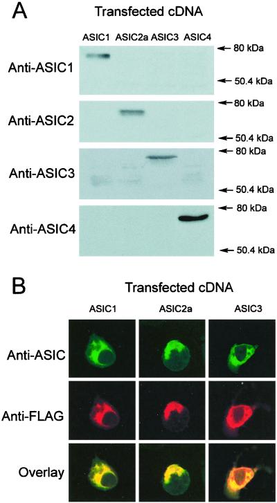
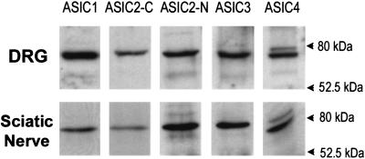
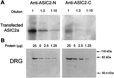
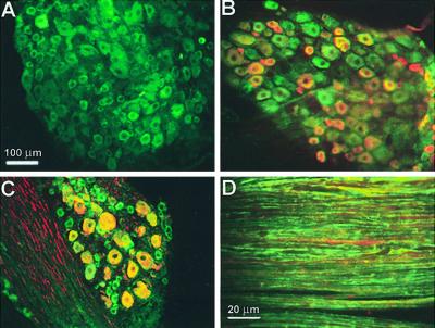
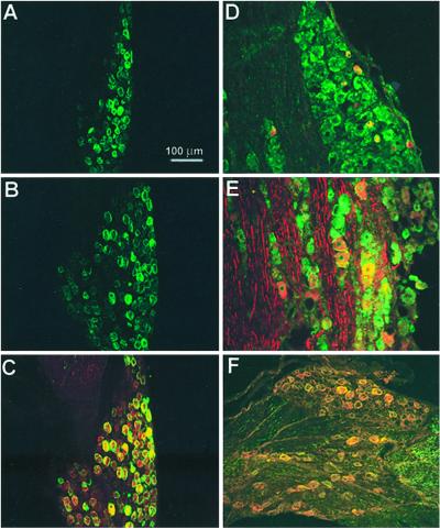
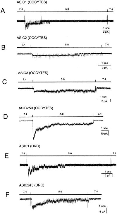
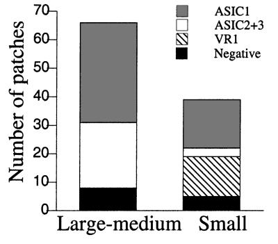
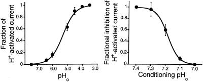
Acid-sensitive ion channels (ASIC) are proton-gated ion channels expressed in neurons of the mammalian central and peripheral nervous systems. The functional role of these channels is still uncertain, but they have been proposed to constitute mechanoreceptors and/or nociceptors. We have raised specific antibodies for ASIC1, ASIC2, ASIC3, and ASIC4 to examine the distribution of these proteins in neurons from dorsal root ganglia (DRG) and to determine their subcellular localization. Western blot analysis demonstrates that all four ASIC proteins are expressed in DRG and sciatic nerve. Immunohistochemical experiments and functional measurements of unitary currents from the ASICs with the patch-clamp technique indicate that ASIC1 localizes to the plasma membrane of small-, medium-, and large-diameter cells, whereas ASIC2 and ASIC3 are preferentially in medium to large cells. Neurons coexpressing ASIC2 and ASIC3 form predominantly heteromeric ASIC2-3 channels. Two spliced forms, ASIC2a and ASIC2b, colocalize in the same population of DRG neurons. Within cells, the ASICs are present mainly on the plasma membrane of the soma and cellular processes. Functional studies indicate that the pH sensitivity for inactivation of ASIC1 is much higher than the one for activation; hence, increases in proton concentration will inactivate the channel. These functional properties and localization in DRG have profound implications for the putative functional roles of ASICs in the nervous system.


Figures

Figure 1
Figure 1
Characterization of ASIC antibodies. (A) Western blots from HEK cells transfected with ASIC-1, -2a, -3, and -4 cDNAs probed with affinity-purified specific antibodies. (B) Immunocytochemistry of HEK cells transfected with ASIC-1, -2, and -3 cDNAs tagged with FLAG epitope. (Top) Confocal images of cells labeled with the corresponding anti-ASIC antibodies (green); (Middle) labeling with monoclonal anti-FLAG antibody (red); (Bottom) the merge of the previous two images (yellow).
Figure 2
Figure 2
Western blots of DRG and sciatic nerve. Analysis of 50 and 80 μg of proteins extracted from DRG and sciatic nerve, respectively, with five different antibodies for: ASIC1, ASIC2-C (recognizes the C terminus of ASIC2a and ASIC2b), anti-ASIC2-N (specific for the N terminus of ASIC2a), ASIC3, and ASIC4.
Figure 3
Figure 3
Apparent affinities of anti-ASIC2-N and anti-ASIC2-C antibodies examined by Western blots. (A) Serial dilutions of lysates from HEK transfected with ASIC2a; (B) serial dilutions of lysates from DRG. Membranes were probed with ASIC2-N or ASIC2-C antibodies.
Figure 4
Figure 4
Immunolocalization of ASIC1 in DRG. (A) Staining with ASIC1 antibody. (B) Double staining with ASIC1 (green) and monoclonal antiperipherin (red). (C) Double staining with ASIC1 (green) and monoclonal antineurofilament 200 (red). (D) Section of sciatic nerve labeled with ASIC1 (green) and monoclonal antineurofilament 200 (red). ASIC1 localizes to small-, medium-, and large-diameter cells and overlaps with peripherin or neurofilament 200. In sciatic nerve ASIC1 stains the plasmalemma of axons.
Figure 5
Figure 5
Immunolocalization of ASIC2 and ASIC3 in DRG. (A) Staining of DRG with ASIC2-N antibody; (B) staining with ASIC2-C; (C) double staining with ASIC2-N (red) and ASIC2-C (green); (D) double staining with ASIC3 (green) and anti-peripherin monoclonal antibody (red) or with (E) antineurofilament 200 monoclonal antibody (red). (F) Double staining with ASIC2-C (green) and ASIC3 (red) antibodies.
Figure 6
Figure 6
Representative examples of acid-activated currents in oocytes injected with different ASIC cRNAs and in freshly isolated neurons from DRG. Recordings were obtained from outside-out patches held at −40 mV. Bars above traces indicate the change in pH0. The spikes in the records are artifacts introduced by the change of solution. Bars indicate the scale for time and current amplitudes. Currents from ASIC1 (A), ASIC2 (B), ASIC3 (C), and ASIC2–3 (D) expressed in oocytes. (E) Currents with characteristics of ASIC1 in small cells from DRG. (F) Currents with characteristics of ASIC2–3 in medium-large DRG cells.
Figure 7
Figure 7
Frequency of ASIC channels in DRG neurons. Stack columns represent the frequency of finding various types of ASICs in outside-out patches from freshly isolated DRG cells. Cells were classified as small or medium-large by visual inspection under the microscope of the patch-clamp setup. Channels were assigned to categories according to the properties of inactivation, single channel kinetics, current sublevels, and block by ruthenium red. The conditions of the experiments were the same as in oocytes.
Figure 8
Figure 8
Dose-response curves of activation and inactivation of ASIC1 by increasing [H+]o. Whole-cell currents from oocytes expressing ASIC1 were recorded with the two-microelectrode voltage-clamp technique. (Left) Currents induced by stepwise decrease in pHo were normalized to the maximal value obtained when the pHo was rapidly changed from 7.4 to 4.0. The solid line represents the fit of the data to the equation IMax = 1/(1 + [(Kd/pHo)N] with values for Kd of 5.3 and N of 1.3. (Right) Currents induced by changing the bath solution from progressively more acidic conditioning pHo values to a constant pHo of 5.0. Data points represent the mean ± SD of at least four oocytes.

Similar articles

Cited by

References

    1. Fyfe G K, Quin A M, Canessa C M. Semin Nephrol. 1998;18:138–151. - PubMed
    1. García-Añoveros J, Derfler B, Neville-Golden J, Hyman B T, Corey D P. Proc Natl Acad Sci USA. 1997;94:1459–1464. - PMC - PubMed
    1. Waldmann R, Champigny G, Bassilana F, Heurteaux C, Lazdunski M. Nature (London) 1997;386:173–177. - PubMed
    1. Chen C, England S, Akopian A N, Wood J N. Proc Natl Acad Sci USA. 1998;95:10240–10245. - PMC - PubMed
    1. Price M P, Snyder P M, Welsh M J. J Biol Chem. 1996;271:7879–7882. - PubMed

Publication types

MeSH terms

LinkOut - more resources