. 2002 Feb 19;99(4):1966-71.
doi: 10.1073/pnas.042689399. Epub 2002 Feb 12.

Alveolar epithelial type I cells contain transport proteins and transport sodium, supporting an active role for type I cells in regulation of lung liquid homeostasis

Affiliations

Alveolar epithelial type I cells contain transport proteins and transport sodium, supporting an active role for type I cells in regulation of lung liquid homeostasis

Meshell D Johnson et al. Proc Natl Acad Sci U S A. .

Abstract

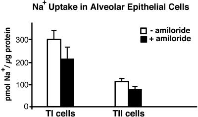
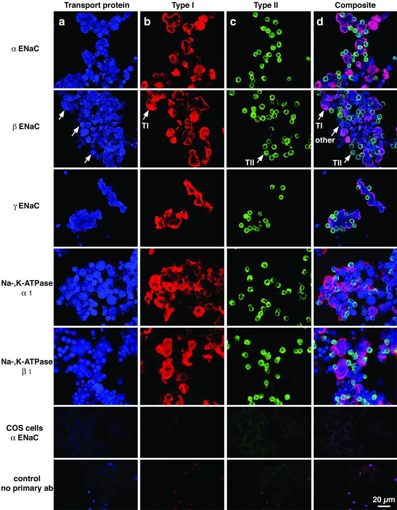
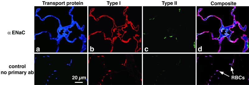
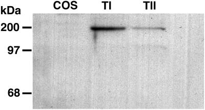
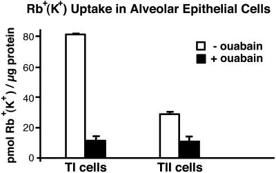
Transport of lung liquid is essential for both normal pulmonary physiologic processes and for resolution of pathologic processes. The large internal surface area of the lung is lined by alveolar epithelial type I (TI) and type II (TII) cells; TI cells line >95% of this surface, TII cells <5%. Fluid transport is regulated by ion transport, with water movement following passively. Current concepts are that TII cells are the main sites of ion transport in the lung. TI cells have been thought to provide only passive barrier, rather than active, functions. Because TI cells line most of the internal surface area of the lung, we hypothesized that TI cells could be important in the regulation of lung liquid homeostasis. We measured both Na(+) and K(+) (Rb(+)) transport in TI cells isolated from adult rat lungs and compared the results to those of concomitant experiments with isolated TII cells. TI cells take up Na(+) in an amiloride-inhibitable fashion, suggesting the presence of Na(+) channels; TI cell Na(+) uptake, per microgram of protein, is approximately 2.5 times that of TII cells. Rb(+) uptake in TI cells was approximately 3 times that in TII cells and was inhibited by 10(-4) M ouabain, the latter observation suggesting that TI cells exhibit Na(+)-, K(+)-ATPase activity. By immunocytochemical methods, TI cells contain all three subunits (alpha, beta, and gamma) of the epithelial sodium channel ENaC and two subunits of Na(+)-, K(+)-ATPase. By Western blot analysis, TI cells contain approximately 3 times the amount of alphaENaC/microg protein of TII cells. Taken together, these studies demonstrate that TI cells not only contain molecular machinery necessary for active ion transport, but also transport ions. These results modify some basic concepts about lung liquid transport, suggesting that TI cells may contribute significantly in maintaining alveolar fluid balance and in resolving airspace edema.


Figures

Figure 1
Figure 1
Uptake of Na+ by TI and TII cells in vitro. Na+ uptake was performed as described in Methods. Data are expressed as mean ± SE, n = 4 for TI cells and n = 6 for TII cells. Na+ uptake in both TI and TII cells was significantly inhibited by amiloride (29% in TI with P < 0.001; 28% in TII with P < 0.03). Na+ uptake in TI cells per microgram of protein was greater than that in TII cells (P < 0.01).
Figure 2
Figure 2
Colocalization of subunits of ENaC and Na+-, K+-ATPase with markers specific for TI and TII cells. The preparations of isolated cells used in these experiments contain not only TI cells and TII cells, but various other lung cells liberated by enzymatic digestion, including airway cells, macrophages, and cells from the vascular and interstitial compartments. Immunocytochemistry was performed as described in Methods. The same field of cells is shown in each row, stained with three antibodies and three different fluorophores specific for each antibody. The colors shown in each column are the result of scanning with the appropriate laser for each fluorophore. In the various columns, one can appreciate staining for (a) antibodies against various channel or pump proteins (blue color); (b) RTI40, a marker for TI cells (red color); and (c) RTII70, a marker for TII cells (green color). Column d shows a composite image, created by superimposition of the three separate images shown in columns a, b, and c. Arrows indicate one representative TI cell, one TII cell, and a cell that is neither a TI nor a TII cell. Every TI and TII cell expressed all of the pump/channel proteins. This fact can be appreciated most easily when one looks in column d at the spectral shifts caused by overlapping red (TI) or green (TII) markers with the blue (pump/channel) protein. In this composite image, colocalization of a transport protein subunit (ENaC or Na+-, K+-ATPase) with either TI or TII cell plasma membrane causes a color shift, indicating colocalization of the protein subunit with TI or TII plasma membranes. For example, combination of blue (subunit) with red (TI) is pinkish purple, whereas a combination of blue (subunit) with green (TII) is bluish green. Specific antibodies are labeled in rows 1–5: row 1, αENaC; row 2, βENaC; row 3, γENaC; row 4, α1-Na+-, K+-ATPase; row 5, β1-Na+-, K+-ATPase; row 6, Cos7 cells stained for αENaC; and row 7, no primary antibody control panel with mixed lung cells.
Figure 3
Figure 3
αENaC is present in TI cells in situ. (Upper) A 2-μ-thick cryostat section of rat lung incubated with primary and secondary antibodies and scanned sequentially as described in Methods. The organization is the same as described for Fig. 2: (a) αENaC (blue); (b) RTI40 (TI cell apical plasma membrane; red); (c) RTII70 (TII cell plasma membrane; green); and (d) computer-generated composite image. (Lower) A 2-μ-thick section prepared without primary antibodies but with all three secondary antibodies. Note that erythrocytes (RBCs) autofluoresce similarly with and without primary antibody.
Figure 4
Figure 4
Western blot for αENaC in Cos, TI, and TII cells. Forty-micrograms of protein from Cos7, TI, or TII cells was added to each lane, and bands were resolved on a 10% Bis-Tris gel, transferred to nitrocellulose paper, and stained with an antibody specific for αENaC as described in Methods. A band at ≈180–200 kDa is seen in TI and TII cell lanes. By PhosphorImager quantitation, TI cells express ≈3 times the amount of αENaC found in TII cells.
Figure 5
Figure 5
Rb+ uptake in freshly isolated TI and TII cells. Rb+ uptake in freshly isolated TI and TII cells at 5 min with and without ouabain (10−4 M). Data are expressed as mean ± SE, n = 2 for each cell type. In T1 cells, ouabain inhibited Rb+ uptake by an average of 85% (P < 0.02)

Similar articles

Cited by

References

    1. Diamond J M. J Membr Biol. 1979;51:195–216. - PubMed
    1. Saumon G, Basset G. J Appl Physiol. 1993;74:1–15. - PubMed
    1. Matalon S, Benos D J, Jackson R M. Am J Physiol. 1996;271:L1–L22. - PubMed
    1. Matalon S, O'Brodovich H. Annu Rev Physiol. 1999;61:627–661. - PubMed
    1. Weibel E R. Morphometry of the Human Lung. Berlin: Springer; 1963.

Publication types

LinkOut - more resources