. 2002 Mar;184(5):1335-9.
doi: 10.1128/JB.184.5.1335-1339.2002.

Trinucleotide GAA repeats dictate pMGA gene expression in Mycoplasma gallisepticum by affecting spacing between flanking regions

Affiliations

Trinucleotide GAA repeats dictate pMGA gene expression in Mycoplasma gallisepticum by affecting spacing between flanking regions

Li Liu et al. J Bacteriol. 2002 Mar.

Abstract

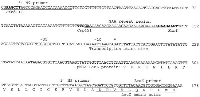
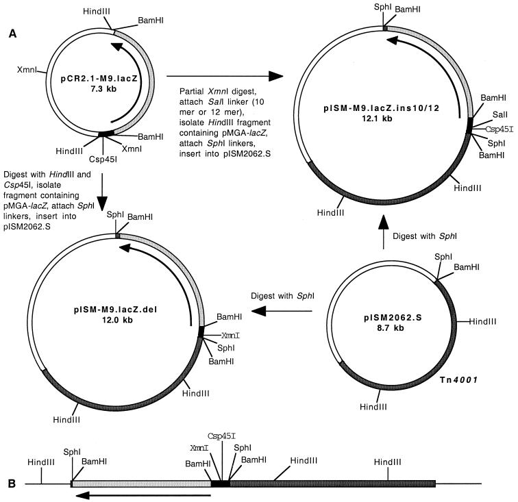
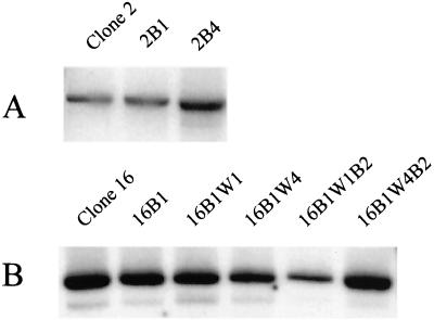
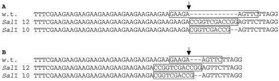
The pMGA genes of the avian respiratory pathogen Mycoplasma gallisepticum encode a family of hemagglutinins that are subject to phase variation. A trinucleotide GAA repeat region is located upstream of the pMGA transcription start site. The length of the repeat region varies at a high frequency due to changes in the number of repeat units. Previous studies have shown that pMGA genes are transcribed when 12 GAA repeats are present but are not transcribed when the number of repeats is not 12. To further analyze the mechanism of gene regulation, the pMGA promoter region was modified either by deleting the nucleotides 5" of the GAA repeats or by inserting linkers of 10 or 12 bp at a position 3" of the repeats. The modified promoter region was fused to a promoterless lacZ gene and transformed into M. gallisepticum by using transposon Tn4001 as a vector. Transformants and successive generations of progeny were analyzed with 5-bromo-4-chloro-3-indolyl-beta-D-galactopyranoside (X-Gal) to monitor beta-galactosidase activity. For the transformants of M. gallisepticum containing the reporter with deletion of nucleotides 5" of the GAA repeats, GAA-dependent pMGA gene regulation was abolished. For the transformants containing the reporter with an addition of 10- or 12-bp linkers, lacZ was expressed only when eight GAA repeats were present. These data indicate that the nucleotides 5" of the GAA repeats as well as the spacing between the GAA repeats and sequences downstream (3") of the repeats are important for pMGA gene expression.


Figures

FIG. 1.
FIG. 1.
Nucleotide sequence of the 5" end region of the unmodified pMGA-lacZ fusion gene. Sequences corresponding to the oligonucleotide primers used for PCR amplification and DNA sequencing are underlined. Also underlined are the GAA repeats, the amino acids encoded by lacZ, and the −10 and −35 regions of the promoter. The putative transcription start site (asterisk) was identified based on sequence similarity to the previously determined start site of pMGA1.1 (5). Restriction enzyme recognition sites are in bold.
FIG. 2.
FIG. 2.
Schematic diagram of the construction of the modified pMGA-lacZ reporters and their insertion into the chromosome of M. gallisepticum. (A) Deletion of nucleotides to generate pISM-M9.lacZ.del and insertion of SalI linkers to generate pISM-M9.lacZ.ins10/12. White regions denote sequences derived from E. coli plasmid vectors. Regions in black, dark gray, and light gray denote the pMGA gene fragment, sequences originating from Tn4001, and lacZ, respectively. Arrows in plasmids show the direction of transcription. (B) A representation of Tn4001 containing pMGA-lacZ inserted into the chromosome of M. gallisepticum. Thin lines denote mycoplasmal chromosomal DNA flanking the transposon. Shaded regions are as in panel A. The leftmost HindIII site is the mycoplasmal HindIII site most proximal to the left end of the transposon. The XmnI site in the pMGA promoter region is replaced with SalI in the transposon of pISM-M9.lacZ.ins10 and pISM-M9.lacZ.ins12. The Csp45I site and upstream pMGA sequences were deleted in the transposon of pISM-M9.lacZ.del.
FIG.3.
FIG.3.
Southern analysis of mycoplasmal genomic DNA digested with HindIII and probed with lacZ. Controls showing that the lacZ probe does not hybridize with DNA from M. gallisepticum strain PG31 that lacks the pMGA-lacZ reporter were previously described (7). (A) Clone 2 is a LacZ transformant of M. gallisepticum transformed with pISM-M9.lacZ.del. Clones 2B1 and 2B4 are LacZ+ progeny of clone 2. (B) Clone 16 is a LacZ transformant of M. gallisepticum transformed with pISM-M9.lacZ.ins.10. The lineage and phenotype of LacZ+ and LacZ derivatives of clone 16 (clones 16B1, 16B1W1, 16B1W4, 16B1W1B2, and 16B1W4B2) are provided in Fig. 4. As determined by comparison to the mobility of radiolabeled HindIII fragments of bacteriophage lambda DNA, the hybridizing DNA fragments from clones 2 and 16 are about 15 and 5 kb, respectively. Similar experiments (data not shown) indicate that transposition of Tn4001 also did not occur during switching of LacZ production in M. gallisepticum transformed with pISM-M9.lacZ.ins.12.
FIG. 4.
FIG. 4.
Pedigree analysis of M. gallisepticum containing the pMGA-lacZ reporter modified by insertion of 10- and 12-bp linkers. For each of the subclones, the percentage of progeny that were scored as blue, lacZ+ colonies is indicated.
FIG. 5.
FIG. 5.
Sequence alignment of the pMGA GAA repeat region of the unmodified promoter (w.t.) and promoters modified by the addition of 10-bp (SalI 10) and 12-bp (SalI 12) linkers. For the unmodified promoter, the XmnI site is boxed, with an arrow showing the site of XmnI cleavage. For the SalI-modified promoters, the SalI linker is boxed. Dashes refer to gaps in the nucleotide sequence introduced to realign the sequences 3" of the XmnI site or linker. (A) Alignment of the unmodified sequence to that of the original SalI-modified sequences that had 12 GAA repeats and gave rise to LacZ transformants; (B) alignment of sequences from LacZ+ clones.
FIG. 6.
FIG. 6.
Schematic illustration of the binding of HAP to sequences upstream and downstream of 12 GAA repeats. Numbers are distances (in nucleotides) from the indicated position to the transcription start site. Restriction enzyme sites: H, HindIII; C, Csp45I; X, XmnI.

References

    1. Baseggio, N., M. D. Glew, P. F. Markham, K. G. Whithear, and G. F. Browning. 1996. Size and genomic location of the pMGA multigene family of Mycoplasma gallisepticum. Microbiology 142:1429-1435. - PubMed
    1. Dybvig, K., and L. L. Voelker. 1996. Molecular biology of mycoplasmas. Annu. Rev. Microbiol. 50:25-57. - PubMed
    1. Glew, M. D., N. Baseggio, P. F. Markham, G. F. Browning, and I. D. Walker. 1998. Expression of the pMGA genes of Mycoplasma gallisepticum is controlled by variation in the GAA trinucleotide repeat lengths within the 5" noncoding regions. Infect. Immun. 66:5833-5841. - PMC - PubMed
    1. Glew, M. D., G. F. Browning, P. F. Markham, and I. D. Walker. 2000. pMGA phenotypic variation in Mycoplasma gallisepticum occurs in vivo and is mediated by trinucleotide repeat length variation. Infect. Immun. 68:6027-6033. - PMC - PubMed
    1. Glew, M. D., P. F. Markham, G. F. Browning, and I. D. Walker. 1995. Expression studies on four members of the pMGA multigene family in Mycoplasma gallisepticum S6. Microbiology 141:3005-3014. - PubMed

Publication types

MeSH terms

Substances

LinkOut - more resources