. 2002 Feb;23(2):243-7.

Astroblastoma: radiologic-pathologic correlation and distinction from ependymoma

Affiliations

Astroblastoma: radiologic-pathologic correlation and distinction from ependymoma

John D Port et al. AJNR Am J Neuroradiol. 2002 Feb.

Abstract

Astroblastoma is a rare primary glial tumor with a characteristic appearance on neuroradiologic images. Typically, astroblastomas are large, lobulated, peripheral, supratentorial, solid, and cystic masses with relatively little associated vasogenic edema and tumor infiltration for their large size. The solid component of the mass has a bubbly appearance and a T2 signal that is isointense to gray matter. Punctate calcifications are often present. Neuroradiologists should be familiar with the characteristic appearance of this tumor.


Figures

F<sc>ig</sc> 1.
Fig 1.
Histopathologic specimens of a well-differentiated and malignant astroblastoma. A, Well-differentiated astroblastomas are composed of astroblastic pseudorosettes, which are elongated tumor cells with broad or tapering processes extending to a central vessel (arrow). B, Malignant astroblastomas contain hypercellular mitotically active regions that often display vascular proliferation or necrosis (arrowheads).
F<sc>ig</sc> 2.
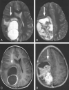
Fig 2.
The classic MR imaging appearance of an astroblastoma in a 3-year-old female patient (patient 5). A and B, Axial T2-weighted images [2500/90/1 (TR/TE/excitations)] depict a large, well-circumscribed, heterogeneous supratentorial mass with peripheral cystic changes (arrows). Note that the solid component of the mass has a heterogeneous bubbly appearance in its center. C and D, Axial postcontrast T1-weighted images (500/20/1) show rim enhancement of the cystic component (arrows) and heterogeneous enhancement of the solid component. In this case, the tumor was malignant.
F<sc>ig</sc> 3.
Fig 3.
The classic CT and MR imaging appearance of an astroblastoma in a 42-year-old female patient (patient 2). A, Axial non-contrast-enhanced CT scan shows punctate calcifications (arrowheads) within the solid portion of the large left temporal lobe mass (arrow). B, Axial non-contrast-enhanced T1-weighted image (500/8/1) shows the solid portion of the tumor (arrow) to be relatively hypointense to gray matter. C, Axial T2-weighted image (2640/98/1) shows the solid portion of the tumor (arrow) as isointense to gray matter. This tumor was well differentiated.
F<sc>ig</sc> 4.
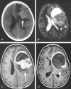
Fig 4.
The classic CT and MR imaging appearance of an astroblastoma in a 5-year-old female patient (patient 4). A, Axial non-contrast-enhanced CT scan shows the solid component of the mass has a slightly higher attenuation than that of the adjacent gray matter (arrow). B, Axial T2-weighted image (7935/115/1) depicts the relative lack of peritumoral T2 hyperintensity (representing vasogenic edema or tumor infiltration or both) given the large tumor size (arrowheads). C and D, Axial postcontrast T1-weighted images (450/18/1) depict intense heterogeneous enhancement of the solid portion (C, arrow) and rim enhancement of the cystic portion (D, arrow). The diagnosis of a malignant astroblastoma was confirmed on the basis of histologic findings.
F<sc>ig</sc> 5.
Fig 5.
MR images of an aggressive astroblastoma in a 15-year-old female patient (patient 6). A, Sagittal T1-weighted image (810/14/1) shows an aggressive mass that has invaded the anterior body of the corpus callosum (arrowhead). B, Axial T2-weighted image (5000/93/1) shows the lesion to have slightly higher T2 signal intensity than that typically seen with an astroblastoma, and the mass appears centered within the anterior horn of the left lateral ventricle (arrow), suggesting the diagnosis of ependymoma. C, Coronal postcontrast T1-weighted image (950/14/1) depicts heterogeneous enhancement of the solid portion (arrowhead) and rim enhancement around the cystic portion (arrow). The tumor was a malignant astroblastoma.
F<sc>ig</sc> 6.
Fig 6.
MR images of a relatively small astroblastoma in a 30-year-old male patient (patient 1). A, Axial T2-weighted image (3466/84/1) shows the solid component of this left frontal lesion has the characteristic bubbly appearance of an astroblastoma (arrow), but a moderate amount of peritumoral T2 hyperintensity associated with the lesion (arrowheads) is present. Furthermore, the cystic component is absent. B, Axial postcontrast T1-weighted image (550/8) shows the typically intense heterogeneous enhancement of the solid component. The overall imaging characteristics were nonspecific; histologic analysis confirmed the diagnosis of a well-differentiated astroblastoma.

References

    1. Pizer BL, Moss T, Oakhill A, Webb D, Coakham HB. Congenital astroblastoma: an immunohistochemical study. Case report. J Neurosurg 1995;83:550–555 - PubMed
    1. Bailey P, Cushing H. Classification of the Tumors of the Glioma Group. Philadelphia, Pa: Lippincott;1926. :83–84; 133–136
    1. Bailey P, Bucy PC. Astroblastomas of the brain. Acta Psychiatr Neurol 1930;5:439–461
    1. Brat DJ, Hirose Y, Cohen KJ, Feuerstein BG, Burger PC. Astroblastoma: clinicopathologic features and chromosomal abnormalities defined by comparative genomic hybridization. Brain Pathol 2000;10:342–352 - PMC - PubMed
    1. Bonnin JM, Rubinstein LJ. Astroblastomas: a pathological study of 23 tumors, with a postoperative follow-up in 13 patients. Neurosurgery 1989;25:6–13 - PubMed