. 2002 Feb 15;21(4):493-503.
doi: 10.1093/emboj/21.4.493.

Regulation of receptor protein-tyrosine phosphatase alpha by oxidative stress

Affiliations

Regulation of receptor protein-tyrosine phosphatase alpha by oxidative stress

Christophe Blanchetot et al. EMBO J. .

Abstract

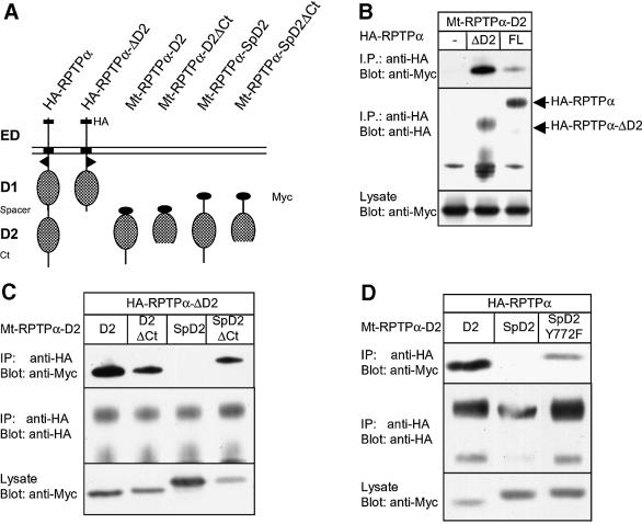
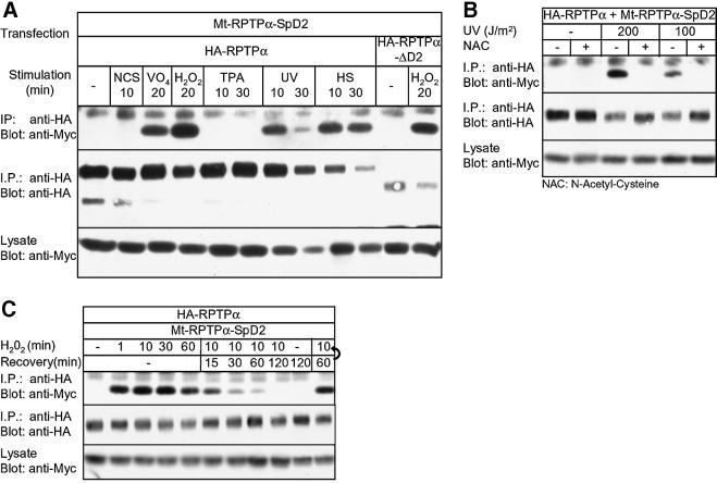
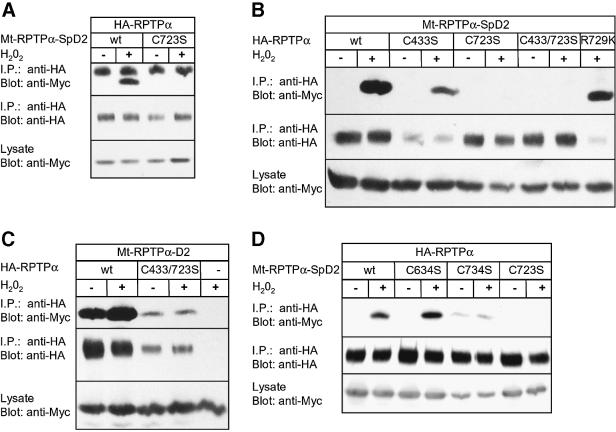
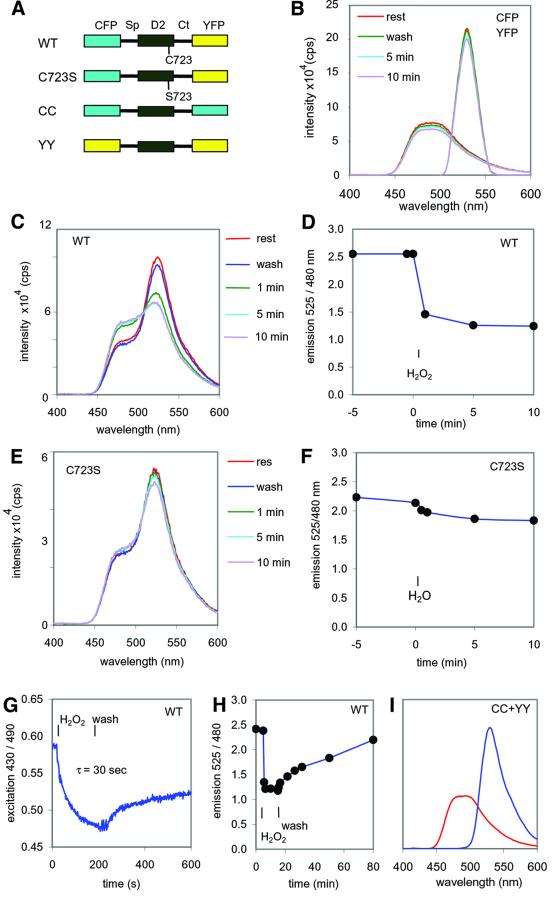
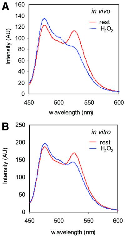
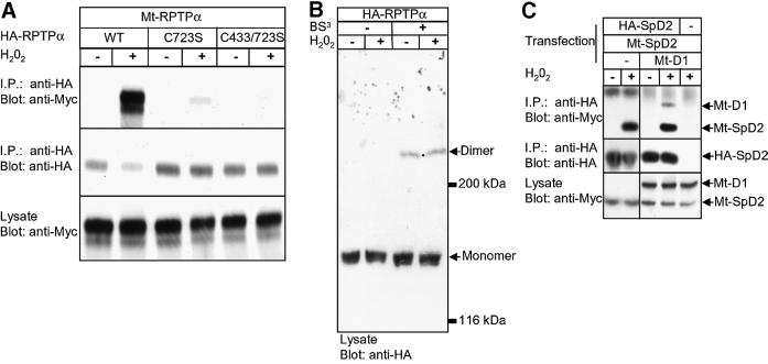
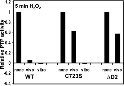
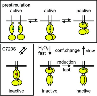
The presence of two protein-tyrosine phosphatase (PTP) domains is a striking feature in most transmembrane receptor PTPs (RPTPs). The function of the generally inactive membrane-distal PTP domain (RPTP-D2) is unknown. Here we report that an intramolecular interaction between the spacer region (Sp) and the C-terminus in RPTPalpha prohibited intermolecular interactions. Interestingly, stress factors such as H(2)O(2), UV and heat shock induced reversible, free radical-dependent, intermolecular interactions between RPTPalpha and RPTPalpha-SpD2, suggesting an inducible switch in conformation and binding. The catalytic site cysteine of RPTPalpha-SpD2, Cys723, was required for the H(2)O(2) effect on RPTPalpha. H(2)O(2) induced a rapid, reversible, Cys723-dependent conformational change in vivo, as detected by fluorescence resonance energy transfer, with cyan fluorescent protein (CFP) and yellow fluorescent protein (YFP) flanking RPTPalpha-SpD2 in a single chimeric protein. Importantly, H(2)O(2) treatment stabilized RPTPalpha dimers, resulting in inactivation. We propose a model in which oxidative stress induces a conformational change in RPTPalpha-D2, leading to stabilization of RPTPalpha dimers, and thus to inhibition of RPTPalpha activity.


Figures

None
Fig. 1. The spacer and the C-terminal region of RPTPα together block binding to RPTPα. (A) Schematic representation of the constructs used here. HA, HA tag; ED, extracellular domain; D1, RPTP-D1; spacer, region between RPTP-D1 and RPTP-D2; D2, RPTP-D2; Ct, C-terminus; Myc, Myc tag. (B–D) Co-immunoprecipitation experiments. 293 cells were transiently co-transfected with the constructs indicated. HA-tagged RPTPα was immunoprecipitated using anti-HA antibodies (12CA5), resolved on SDS–PAGE gels and blotted. The gels were probed with anti-Myc antibody (9E10; top panels) and anti-HA (12CA5; middle panels). Expression of Myc-tagged RPTPα-D2s (Mt-RPTPα-D2s) in the lysates was monitored in the bottom panels. (B) Co-transfection of Mt-RPTPα-D2 alone (–), with full-length HA-tagged RPTPα (FL) or mutant RPTPα lacking the second domain (ΔD2). (C) Co-transfection of HA-RPTPα-ΔD2 with various Mt-RPTPα-D2 constructs [depicted in (A)]. (D) Co-transfection of full-length HA-RPTPα with Mt-RPTPα-D2, -SpD2 or SpD2Y772F.
None
Fig. 2. Binding of RPTPα-SpD2 to RPTPα is induced by oxidative stress and is reversible. (A) 293 cells were co-transfected with HA-RPTPα or HA-RPTPα-ΔD2 and Mt-RPTPα-SpD2. After stimulation with (final concentration) 10% newborn calf serum (NCS), pervanadate (VO4; 1 mM VO4 + 1 mM H2O2), 1 mM H2O2, 50 ng/ml TPA, UV (200 J/m2) or heat shock (HS; 42°C) for the time indicated, HA-RPTPα was immuno precipitated. After separation on SDS–PAGE and blotting of the gel, the blot was probed with anti-Myc (top panel) and anti-HA (middle panel) antibodies. Equal expression of the Myc-tagged RPTP-D2s was monitored in the lower panel. (B) 293 cells were transiently transfected with HA-RPTPα and Mt-RPTPα-SpD2, pre-treated (+) or not (–) with 10 mM NAC for 16 h and subjected to 100 or 200 J/m2 UV radiation, and left to recover for 10 min. Immunoprecipitation and immunoblotting were as in (A). (C) Transiently transfected 293 cells were treated with H2O2 (1 mM) for the time indicated. Alternatively, after 10 min of stimulation with H2O2, the medium was replaced with fresh pre-warmed medium for the time indicated (Recovery). Note that media change by itself did not affect H2O2-induced binding (last lane). Immunoprecipitation and immunoblotting were as in (A).
None
Fig. 3. H2O2-induced binding of RPTPα-SpD2 to RPTPα is dependent on catalytic site Cys723. (A) 293 cells were co-transfected with HA-RPTPα and wild-type Mt-RPTPα-SpD2 (wt) or mutant Mt-RPTPα-SpD2 with Cys723 mutated to Ser (C723S). The cells were treated with H2O2 (1 mM, 15 min) (+) or left untreated (–). HA-RPTPα was immunoprecipitated, separated on SDS–PAGE gels and blotted. The blot was probed with anti-Myc (top panel) and anti-HA (middle panel) antibodies. Expression of the Mt-RPTP-D2s in the lysate was monitored in the lower panel. (B) Mt-RPTPα-SpD2 was co-transfected in 293 cells with full-length wild-type HA-RPTPα (wt) or mutants, as indicated. The cells were stimulated with H2O2 (1 mM, 15 min). Immunoprecipitation and immunoblotting for (B–D) were carried out as in (A). (C) Mt-RPTPα-SpD2 was co-transfected with full-length HA-RPTPα (wt), HA-RPTPα-C433S/C723S or PSG5 (–) and stimulated with H2O2 (1 mM, 15 min). (D) 293 cells were transiently co-transfected with HA-RPTPα and various Mt-RPTPα-SpD2 mutants with Cys to Ser mutations, as indicated, and treated with H2O2 (1 mM, 5 min).
None
Fig. 4. H2O2-induced conformational change in RPTPα-D2. (A) Schematic representation of the constructs used here: CFP–RPTPα-SpD2–YFP2.1 (WT), CFP–RPTPα-SpD2-C723S–YFP2.1 (C723S), CFP–RPTPα-SpD2–CFP (CC) and YFP–RPTPα-SpD2–YFP (YY). Sp, spacer region; Ct, C-terminus. (B) The spectral properties of a single 293 cell, transfected with CFP alone (excitation at 430 nm, emission maximum at 480 nm), or YFP2.1 alone (excitation at 490 nm, emission maximum at 525 nm), before (rest) and after a wash with fresh medium (wash) and during incubation in medium containing 1 mM H2O2 for the periods of time indicated. (C) Emission spectra of a single transfected 293 human embryonic kidney cell (excitation 430 nm), expressing wild-type CFP–RPTPα-SpD2–YFP2.1 (WT). H2O2 treatment (1 mM) as indicated. (D) Time course of the ratio of the emission intensities at 525 and 480 nm from (C). (E) Emission spectra as in (C) of a single cell expressing mutant CFP–RPTPα-SpD2-C723S–YFP2.1 (C723S). (F) Time course of the ratio of the emission intensities at 525 and 480 nm from (E). (G) Real-time FRET analysis was performed on a single cell expressing CFP–RPTPα-SpD2–YFP2.1 (WT) by continuous dual excitation wavelength measurements [excitation at 430 and 490 nm, emission at 535(30) nm; for details see Tertoolen et al., 2001]. Treatment with 1 mM H2O2 and subsequent recovery were followed. The half-time of decay was calculated (τ = 30 s). (H) A single cell expressing CFP–RPTPα-SpD2–YFP2.1 (WT) was treated with 1 mM H2O2 for 10 min, the medium was replaced by regular medium and emission spectra were obtained at regular intervals. The emission ratio at 525 and 480 nm is plotted against time. (I) Emission spectra of a single cell expressing both CC and YY (excitation at 430 nm, red trace; and to control for YFP expression, excitation at 490 nm, blue trace). All single-cell measurements were repeated at least three times. Representative experiments are depicted here.
None
Fig. 5. The effect of H2O2 on RPTPα-D2 is direct. (A) 293 cells were transfected with wild-type CFP–RPTPα-SpD2–YFP2.1 and lysed in cell lysis buffer. Emission spectra were determined for these lysates (excitation 430 nm). The cells had been left untreated (rest) or were stimulated with 1 mM H2O2 for 5 min prior to lysis (in vivo). (B) As in (A), emission spectra were determined for cell lysates. The cell lysates were left untreated (rest) or treated with 1 mM H2O2 (in vitro). The experiments were repeated at least three times and representative measurements are depicted here.
None
Fig. 6. Oxidative stress stabilized RPTPα dimerization. (A) 293 cells were co-transfected with HA-RPTPα full length [wild-type (WT) or with the catalytic site cysteine single mutant (C723S) or double mutant (C433/C723S)] and Myc-tagged full-length RPTPα (Mt-RPTPα). Cells were treated with H2O2 (1 mM, 10 min) (+) or left untreated (–). After anti-HA immunoprecipitation and immunoblotting, the blot was probed with anti-Myc (top panel) and anti-HA (middle panel) antibodies. Equal expression of the Mt-RPTPα in the lysate was monitored in the lower panel. (B) 293 cells were transfected with HA-RPTPα, treated with H2O2 (1 mM, 10 min) (+) or left untreated. Membrane proteins were cross-linked using the non-cell-permeable cross-linker BS3. An immunoblot of whole-cell lysates is shown with the position of RPTPα monomers and dimers indicated. (C) HA-RPTPα-SpD2 was co-transfected with Mt-RPTPα-D1, Mt-RPTPα-D2 or both. H2O2 treatment (10 min, 1 mM), immunoblotting of anti-HA tag immunoprecipitates and lysates were performed as indicated.
None
Fig. 7. Oxidative stress-induced change in RPTPα activity. HA-RPTPα wild type (WT), HA-RPTPα-C723S (C723S) or HA-RPTPα-ΔD2 (ΔD2), lacking D2, were expressed in 293 cells. Cells were stimulated with H2O2 (1 mM, 5 min) (vivo), HA-RPTPα proteins were immunoprecipitated and PTP activity was assayed using pNPP as a substrate. Alternatively, H2O2 (1 mM) was added after immunoprecipitation of RPTPα from unstimulated cells (vitro). The activity was corrected for RPTPα expression.
None
Fig. 8. A model for regulation of RPTPα by oxidative stress. Under normal conditions, RPTPα monomers and dimers are in equilibrium (pre-stimulation). Whether the dimers are active or inactive depends on their rotational coupling. Oxidative stress (e.g. H2O2) leads to rapid inactivation by oxidation of the catalytic site cysteine in D1 (green to red). Oxidative stress also induces a conformational change in D2, which may drive the formation of stabilized dimers, perhaps through D2–D2 interactions. The stabilized dimers are completely inactive. Mutant RPTPα-C723S lacks a conformational change, does not form stabilized dimers and is not inactivated completely following oxidative stress. After reduction, RPTPα slowly returns to the pre-stimulation state following a conformational change in D2. See text for details.

References

    1. Aricescu A.R., Fulga,T.A., Cismasiu,V., Goody,R.S. and Szedlacsek,S.E. (2001) Intramolecular interactions in protein tyrosine phosphatase RPTPµ: kinetic evidence. Biochem. Biophys. Res. Commun., 280, 319–327. - PubMed
    1. Bae Y.S., Kang,S.W., Seo,M.S., Baines,I.C., Tekle,E., Chock,P.B. and Rhee,S.G. (1997) Epidermal growth factor (EGF)-induced generation of hydrogen peroxide. Role in EGF receptor-mediated tyrosine phosphorylation. J. Biol. Chem., 272, 217–221. - PubMed
    1. Bastiaens P.I. and Pepperkok,R. (2000) Observing proteins in their natural habitat: the living cell. Trends Biochem. Sci., 25, 631–637. - PubMed
    1. Bilwes A.M., den Hertog,J., Hunter,T. and Noel,J.P. (1996) Structural basis for inhibition of receptor protein-tyrosine phosphatase-α by dimerization. Nature, 382, 555–559. - PubMed
    1. Blanchetot C. and den Hertog,J. (2000) Multiple interactions between receptor protein-tyrosine phosphatase (RPTP) α and membrane-distal protein-tyrosine phosphatase domains of various RPTPs. J. Biol. Chem., 275, 12446–12452. - PubMed

Publication types