. 2002 Feb 15;21(4):560-71.
doi: 10.1093/emboj/21.4.560.

Distinct mechanisms of internalization of Neisseria gonorrhoeae by members of the CEACAM receptor family involving Rac1- and Cdc42-dependent and -independent pathways

Affiliations

Distinct mechanisms of internalization of Neisseria gonorrhoeae by members of the CEACAM receptor family involving Rac1- and Cdc42-dependent and -independent pathways

Oliver Billker et al. EMBO J. .

Abstract

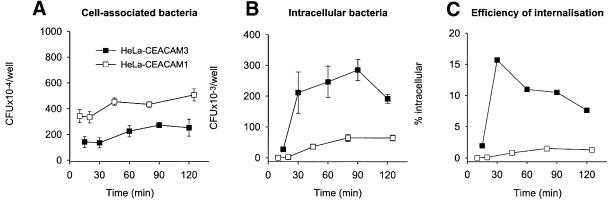
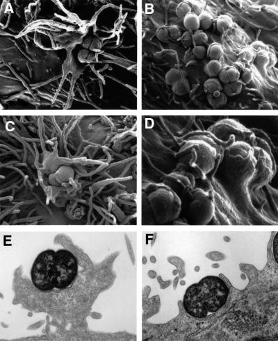
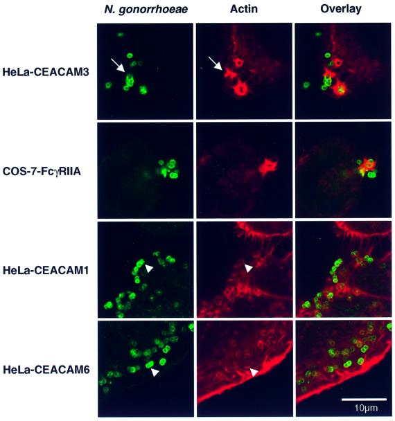
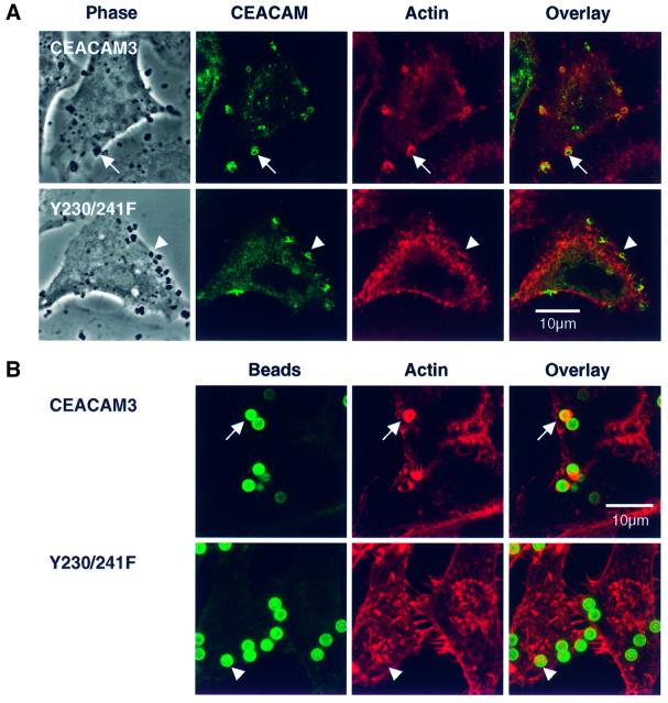
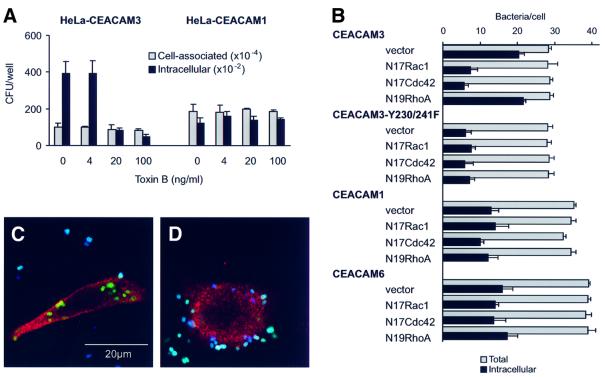
Opa adhesins of pathogenic Neisseria species target four members of the human carcinoembryonic antigen-related cellular adhesion molecule (CEACAM) family. CEACAM receptors mediate opsonization-independent phagocytosis of Neisseria gonorrhoeae by human granulocytes and each receptor individually can mediate gonococcal invasion of epithelial cells. We show here that gonococcal internalization occurs by distinct mechanisms depending on the CEACAM receptor expressed. For the invasion of epithelial cell lines via CEACAM1 and CEACAM6, a pathogen-directed reorganization of the actin cytoskeleton is not required. In marked contrast, ligation of CEACAM3 triggers a dramatic but localized reorganization of the host cell surface leading to highly efficient engulfment of bacteria in a process regulated by the small GTPases Rac1 and Cdc42, but not Rho. Two tyrosine residues of a cytoplasmic immune receptor tyrosine-based activating motif of CEACAM3 are essential for the induction of phagocytic actin structures and subsequent gonococcal internalization. The granulocyte-specific CEACAM3 receptor has properties of a single chain phagocytic receptor and may thus contribute to innate immunity by the elimination of Neisseria and other CEACAM-binding pathogens that colonize human mucosal surfaces.


Figures

None
Fig. 1. Different kinetics of internalization of N.gonorrhoeae N313 (Opa57) by HeLa cell lines expressing either CEACAM3 or CEACAM1. Cells were infected with a multiplicity of infection (m.o.i.) of 30 bacteria per cell. (A) Total cell-associated bacteria. (B) Gentamycin-resistant (i.e. intracellular, viable) bacteria. (C) Rate of internalization shown as the percentage of gentamycin-resistant bacteria calculated from data shown in (A) and (B). Data shown in (A) and (B) are mean results ± SD of triplicate samples for each time point. Data are representative of three independently performed experiments using either N313 (Opa57) or N309 (Opa52).
None
Fig. 2. Neisseria gonorrhoeae N313 (Opa57) triggers different ultrastructural modifications of the cell surface in HeLa lines, depending on the expression of either CEACAM3 or CEACAM1. Subconfluent monolayers were infected with an m.o.i. of 100 for 30 min at 37°C. (AD) SEMs of infected cells showing large cellular protrusions extending from the cell surface of HeLa-CEACAM3 (A and C) and tightly fitting pseudopods enclosing bacteria that adhere to HeLa-CEACAM1 (B and D). (D) shows a detail of (B). Interactions between N313 and a CEACAM6-expressing HeLa cell line were indistinguishable at the ultrastructural level from those with HeLa-CEACAM1 (not shown). (E and F) TEMs of infected HeLa-CEACAM3 (E) and HeLa-CEACAM1 (F). Images are representative of three independent experiments.
None
Fig. 3. Confocal sections showing adherent gonococci and the distinct F-actin structures they trigger in different epithelial cell lines. HeLa lines expressing recombinant CEACAM receptors were infected with N.gonorrhoeae N313 (Opa57), whereas transfected COS-7 cells expressing human FcγRIIa were incubated with immunoglobulin-opsonized N302 (Opa). After 20 min, cells were fixed and processed for indirect immunofluorescence staining of gonococci (green). F-actin was visualized by staining with TRITC–phalloidin (red). F-actin structures induced by the interaction of Opa-expressing bacteria with HeLa-CEACAM3 were highly reminiscent of the phagocytic actin structures triggered by the FcγRIIa receptor. This similarity extended to the formation of distinct phagocytic cups (arrow). In contrast, most Opa-expressing gonococci adhering to CEACAM1- and CEACAM6-expressing cells were only associated with fine rings of F-actin (arrowheads). Images are representative of three or more independent experiments.
None
Fig. 4. Cytochalasin D differentially inhibits CEACAM3-mediated internalization. Following a 30 min pre-incubation period with either cytochalasin D or carrier, HeLa cell lines were infected with N313 (Opa57). After 30 min, total cell-associated bacteria and gentamycin-protected bacteria were quantitated by dilution plating. Black bars show efficient inhibition of gonococcal internalization by HeLa-CEACAM3 (A; black bars) but not by HeLa-CEACAM1 (B), whereas gonococcal adherence (grey bars) to either cell line was not affected by the inhibitor.
None
Fig. 5. Critical role of tyrosine residues in the cytoplasmic receptor domain of CEACAM3 for phagocytic uptake of Opa-expressing gonococci. (A) Cytoplasmic domain mutants and natural splice variants of CEACAM3. (B) Gentamycin protection assay to determine the phagocytic activity of CEACAM3 receptor mutants and splice variants. COS-7 cells expressing the receptor variants shown in (A) or vector transfected control cells were infected with N.gonorrhoeae N313 (Opa57) or N302 (Opa) for 30 min before quantitation of cell-associated and gentamycin-protected bacteria.
None
Fig. 6. The formation of phagocytic F-actin structures by CEACAM3 is triggered by receptor aggregation and critically depends on the tyrosine residues of the cytoplasmic receptor domain. (A) Confocal sections through COS-7 cells transiently transfected to express either CEACAM3 or the double mutant Y230/241F and infected with N313 (Opa57) for 20 min. Phase contrast shows cells and adhering bacteria. Immunofluorescence labelling of CEACAM receptors (green) reveals strong aggregation of both receptors at sites of bacterial adherence. Phagocytic F-actin structures as visualized by TRITC–phalloidin (red) are present in cells expressing wild-type CEACAM3 (arrows) but absent from cells expressing the Y230/241F double mutant (arrowheads). (B) Magnetic beads coated with the D14HD11 mouse monoclonal antibody against CEACAM receptors were incubated with HeLa cell lines for 20 min at 37°C at a ratio of five beads per cell. Receptor aggregation by anti-CEACAM beads is sufficient to induce phagocytic actin rearrangements in cells expressing CEACAM3 (arrows) but not in cells producing the Y230/241F double mutant. Beads were visualized in fixed and permeabilized samples by a dichlorotriazinylamino fluorescein (DTAF)-conjugated secondary antibody against the coating mouse monoclonal antibody.
None
Fig. 7. Different roles for Rho-family GTPases in neisserial internalization via different CEACAM receptors. (A) Gentamycin protection assay of HeLa cell lines infected with N.gonorrhoeae N313 (Opa57) for 30 min at 37°C showing differential inhibition of internalization via CEACAM3 by toxin B. (B) Effects of dominant-interfering GTPase constructs on neisserial internalization by COS-7 cells expressing different CEACAM receptors. Intracellular and extracellular bacteria were determined microscopically for 20 cells in each sample. The bars show the mean percentages of intracellular bacteria and standard deviations of three replicate samples from a single experiment. (C and D) Illustration of the microscopic readout of co-transfection experiments. Confocal sections of COS-7 cells are shown that were first co-transfected with constructs encoding CEACAM3 and either dominant-interfering N19RhoA (C) or N17Cdc42 (D) and then infected with FITC-labelled N313 (Opa57) before fixation and processing for immunofluorescence microscopy. Intracellular bacteria appear green because in non-permeabilized cells they are not accessible to a specific rabbit immune serum and Cy5-conjugated secondary antibody used to label extracellular bacteria, which appear white or blue. Expression of the dominant-interfering GTPase constructs is shown by immunofluorescence labelling against the myc epitope (red) after permeabilization of cells.
None
Fig. 8. Model for CEACAM3 signalling leading to internalization of Neisseria. Following cell contact, CEACAM3-binding Opa variants recruit receptor molecules to the site of adherence. The four CEACAM3-binding Opa variants of the N.gonorrhoeae strain MS11 are shown as an example. Receptor ligation results in phosphorylation of tyrosine residues in the cytoplasmic ITAM motif of the receptor through kinases of the Src family. In granulocytes, Syk kinase may have a key function through its ability to interact specifically with phosphorylated ITAM motifs. However, in epithelial cells that lack Syk, this kinase is not essential. Downstream of receptor activation, the small GTPases Rac and Cdc42 regulate phagocytosis by initiating F-actin assembly, presumably through their downstream effectors such as WASP and the Arp2/3 complex.

Similar articles

Cited by

References

    1. Bos M.P., Grunert,F. and Belland,R.J. (1997) Differential recognition of members of the carcinoembryonic antigen family by Opa variants of Neisseria gonorrhoeae. Infect. Immun., 65, 2353–2361. - PMC - PubMed
    1. Bos M.P., Hogan,D. and Belland,R.J. (1999) Homologue scanning mutagenesis reveals CD66 receptor residues required for neisserial Opa protein binding. J. Exp. Med., 190, 331–340. - PMC - PubMed
    1. Brummer J., Neumaier,M., Gopfert,C. and Wagener,C. (1995) Association of pp60c-src with biliary glycoprotein (CD66a), an adhesion molecule of the carcinoembryonic antigen family downregulated in colorectal carcinomas. Oncogene, 11, 1649–1655. - PubMed
    1. Cambier J.C. (1995) Antigen and Fc receptor signaling. The awesome power of the immunoreceptor tyrosine-based activation motif (ITAM). J. Immunol., 155, 3281–3285. - PubMed
    1. Caron E. and Hall,A. (1998) Identification of two distinct mechanisms of phagocytosis controlled by different Rho GTPases. Science, 282, 1717–1721. - PubMed

Publication types