. 2002 Mar;11(3):659-68.
doi: 10.1110/ps.20402.

The distinctive functions of the two structural calcium atoms in bovine pancreatic deoxyribonuclease

Affiliations

The distinctive functions of the two structural calcium atoms in bovine pancreatic deoxyribonuclease

Ching-Ying Chen et al. Protein Sci. 2002 Mar.

Abstract

The two amino acid residues, Asp 99 and Asp 201, involved in the coordination of the two calcium atoms in the X-ray structure of bovine pancreatic (bp) DNase, were individually changed by site-directed mutagenesis. The two altered proteins, brDNase(D99A) and brDNase(D201A) were expressed in Escherichia coli and purified by anion exchange chromatography. Equilibrium dialysis showed that mutation destroyed one Ca(2+)-binding site each in brDNase(D99A) and brDNase(D201A). Compared with bpDNase, the Vmax value for brDNase(D99A) remained unchanged and that for brDNase(D201A) was decreased, whereas the K(m) values for the two variants were increased two- to threefold when the DNA hydrolytic hyperchromicity assay was used. Like bpDNase, brDNase(D99A) was able to make double scission on duplex DNA with Mg(2+) plus Ca(2+) and was effectively protected by Ca(2+) from the trypsin inactivation. But under the same conditions, brDNase(D201A) lost the double-scission ability and was not protected by Ca(2+). Nevertheless, the two variant proteins retained the characteristics of the Ca(2+)-induced conformational changes and the Ca(2+) protection against the beta-mercaptoethanol disruption of the essential disulfide bond, suggesting that other weaker Ca(2+)-binding sites not found in the X-ray structure were responsible for these properties. Therefore, the two structural calcium atoms are not for maintaining the overall conformation of the active DNase, as it has been indicated in the X-ray analysis, but rather play the role in the fine-tuning of the DNase activity.


Figures
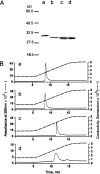
Fig. 1.
Fig. 1.
Homogeneity of the purified proteins. (A) SDS-PAGE of the purified DNases. Lanes a–d were bpDNase, brDNase, brDNase(D99A), and brDNase(D201A), respectively. Because of glycosylation, the native bpDNase is ∼2 kD larger than all of the recombinant proteins. (B) Chromatography of the purified DNases on anion exchanger. The sample was loaded on a Mini Q column, pre-equilibrated with Buffer A (20 mM Tris-HCl at pH 7.5). The flow rate was 0.5 mL/min and the sample was eluted with a linear gradient from 0 to 100% Buffer B (15 mM CaCl2 in Buffer A) for 10 min. The AKTA Purifier System of Amersham Pharmacia Biotech was used for chromatography. (a) bpDNase; (b) brDNase; (c) brDNase(D99A); (d) brDNase(D201A).
Fig. 2.
Fig. 2.
The Scatchard plots for Ca2+ binding. The lines as well as the Kd and n values were obtained by the linear regression analysis based on SigmaPlot for Windows Version 5.00. (Line a) bpDNase, Kd = 1.0 × 10⁻⁵ M, n = 2.6; (line b) brDNase(D99A), Kd = 0.3 × 10⁻⁵ M, n = 1.0; (line c) brDNase(D201A), Kd = 1.3 × 10⁻⁵ M, n = 1.9.
Fig. 3.
Fig. 3.
The modes of duplex DNA scission. The reaction mixture (50 μL) contained 100 μg/mL bovine serum albumin and 140 μg/mL plasmid pCRII DNA with 5 mM CaCl2 or 0.1 mM EGTA in 0.05 M Tris-HCl (pH 7.0), 10 mM MgCl2. Hydrolysis was at 25°C and began after addition of the enzyme. At selected time intervals, 5-μL aliquots of the reaction mixture were quenched with 25 mM EDTA, 6% glycerol, xylene cyanol, and bromphenol blue. Samples were loaded onto 1% agarose gels in the Tris-acetate-EDTA buffer (pH 8.0) containing 0.25 μg/mL of ethidium bromide and a voltage of 8.5 V/cm was applied. (M) DNA molecular weight makers; I, II and III indicate the relaxed, linear, and supercoiled plasmid DNA, respectively.
Fig. 4.
Fig. 4.
Calcium protection against trypsin inactivation. (A) The inactivation kinetics. The final mixture (150 μL) containing 33–133 μg/mL DNase and 3.3–13.3 μg/mL trypsin in 50 mM Tris-HCl (pH 8.0), with 5 mM EDTA (A,a) or 10 mM CaCl2 (A,b), was incubated at 25°C. At selected time intervals, 10-μL aliquots were removed for DNase activity assays. (•) bpDNase; (▾) brDNase(D99A); (○) brDNase(D201A). (B) SDS-PAGE of the trypsin-treated samples. The protein (0.9 μg) in 15 μL of 50 mM Tris-HCl (pH 8.0), was incubated for 10 min with the combinations of 0.09 μg of trypsin, 10 mM Ca2+, or 10 mM EDTA. Prior to loading, all samples were treated with β-mercaptoethanol. Gel was stained with Coomassie blue. The arrows indicate the N-terminal fragments of bpDNase (lane 3), brDNase(D99A) (lane 6) and brDNase(D201A) (lanes 8,9). The C-terminal fragment is not shown.
Fig. 5.
Fig. 5.
The X-ray structure and sequence homology of Sites I and II. (A) Site I with bound calcium. The calcium atom is coordinated by the oxygens of Asp 201, Thr 203, Thr 205, and Thr 207. The loop shown by light ribbon consists of the amino acid residues between Cys 173 and Cys 209, which form the essential disulfide. (B) Site II with bound calcium. The calcium atom is coordinated by the oxygens of Asp 99, Asp 107, Phe 109, and Glu 112. The arrow points toward the flexible loop, GCESCGN. (C) Sequence homology within the motifs of Sites I and II. The alignment for human DNase I, DNase X, DNase γ, and DNAS1L2 was from Shiokawa and Tanuma (2001) and that for bovine and fish DNase I was from Hsiao et al. (1997).
Fig. 6.
Fig. 6.
Effect of Ca2+ on the β-mercaptoethanol inactivation of DNase. The reaction mixture containing 50 mM β-mercaptoethanol in 50 mM Tris-HCl (pH 8.0), with varied amounts of Ca2+ was incubated at 25°C for 15 min and then an equimolar amount of iodoacetamide was added to stop the reaction prior to assay for DNase activities. Samples used, (a) bpDNase; (b) rbDNase(D99A); (c) rbDNase(D201A).
Fig. 7.
Fig. 7.
Calcium-induced spectral changes. (A) The fluorescence spectra in response to Ca2+. (a) bpDNase, 5.2 μg/mL; (b) brDNase(D99A), 7.5 μg/mL; (c) brDNase(D201), 7 μg/mL. The tracings from bottom to top in each panel represent the addition of CaCl2 to the final concentrations of 1 × 10⁻⁷, 10−6, 10−5, 10−4, 10−3, to 5 × 10⁻² M in a and 1 × 10⁻⁷, 2 × 10⁻⁵ and 5 × 10⁻² M in b and c. (B) The Ca2+-induced UV difference spectra. (a) bpDNase, 0.22 mg/mL; (b) brDNase(D99A), 0.25 mg/mL; (c) brDNase(D201A), 0.29 mg/mL.
Fig. 8.
Fig. 8.
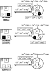
The reaction schemes illustrating the conformational changes, metal bindings, and metal ion-DNA hydrolysis rates for the native and the two variant DNases. I, II, and III represent Sites I, II, and III for the metal ion binding, respectively. C is the catalytic site. CSE refers to as the Ca2+ synergistic effect. Squares and circles represent the inactive and the active conformations of DNases, respectively. The dark areas indicate the impaired sites.

References

    1. Bradord, M.M. 1976. A rapid and sensitive method for the quantitation of microgram quantities of protein utilizing the principle of protein-dye binding. Anal. Biochem. 72 248–254. - PubMed
    1. Campbell, V.W. and Jackson, D.A. 1980. The effect of divalent cations on the mode of action of DNase I. J. Biol. Chem. 255 3726–3735. - PubMed
    1. Chen, C.Y., Lu, S.C., and Liao, T.H. 1998. Cloning, sequencing and expression in Escherichia coli of a DNA encoding bovine pancreatic deoxyribonuclease I: Purification and characterization of the recombinant enzyme. Gene 206181–184. - PubMed
    1. Corns, C.M. and Ludman, C.J. 1987. Some observations on the nature of the calcium-cresolphthalein complexon reaction and its relevance to the clinical laboratory. Ann. Clin. Bioche. 24 345–351. - PubMed
    1. Drayer, A.L., Meima, M.E., Derks, M.W., Tuik, R., and van Haastert, P.J. 1995. Mutation of an EF-hand Ca (2+)-binding motif in phospholipase C of Dictyostelium discoideum: Inhibition of activity but no effect on Ca (2+)-dependence. Biochem. J. 311 505–510. - PMC - PubMed

MeSH terms

LinkOut - more resources