Review
. 2002 Jan;105(1):9-19.
doi: 10.1046/j.0019-2805.2001.01341.x.

IgG4 breaking the rules

Affiliations
Review

IgG4 breaking the rules

Rob C Aalberse et al. Immunology. 2002 Jan.

Abstract

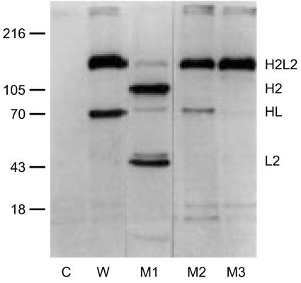
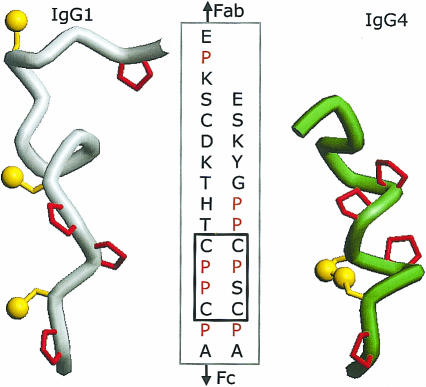
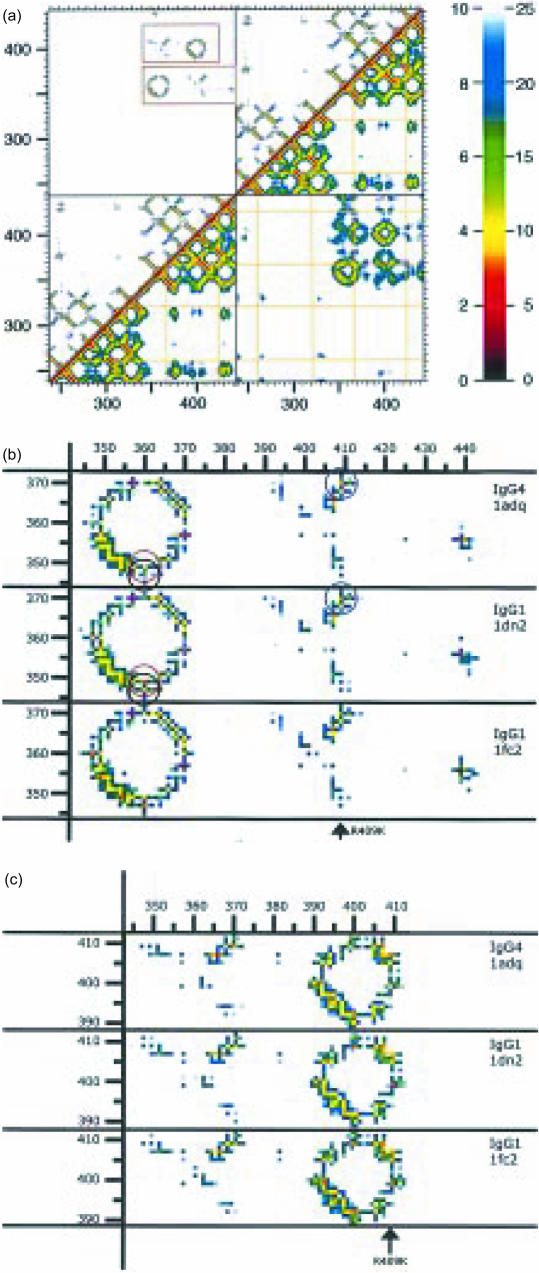
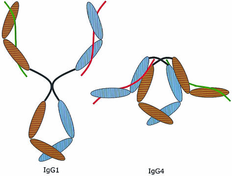
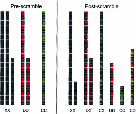
Immunoglobulin G4 (IgG4) antibodies have been known for some time to be functionally monovalent. Recently, the structural basis for this monovalency has been elucidated: the in vivo exchange of IgG half-molecules (one H-plus one L-chain) among IgG4. This process results in bispecific antibodies that in most situations will behave as functionally monovalent antibodies. The structural basis for the abnormal behaviour of IgG4 seems to be largely the result of a single amino acid change relative to human IgG1: the change of a proline in core hinge of IgG1 to serine. This results in a marked shift in the equilibrium between interchain disulphide bridges and intrachain disulphide bridges, which for IgG4 results in 25-75% absence of a covalent interaction between the H-chains. Because of strong non-covalent interactions between the CH3 domains (and possibly also between the CH1 domain and the trans-CH2 domain) IgG4 is a stable four-chain molecule and does not easily exchange half-molecules under standard physiological conditions in vitro. We postulate that the exchange is catalysed in vivo by protein disulphide isomerase (PDI) and/or FcRn (the major histocompatibility complex (MHC)-related Fc receptor) during transit of IgG4 in the endosomal pathway in endothelial cells. Because IgG4 is predominantly expressed under conditions of chronic antigen exposure, the biological relevance of this exchange of half-molecules is that it generates antibodies that are unable to form large immune complexes and therefore have a low potential for inducing immune inflammation. In contrast to monovalent immunoglobulin fragments, these scrambled immunoglobulins have a normal half-life. The significance of the ensuing bispecificity needs further evaluation, because this will be relevant only in situations where high IgG4 responses are found to two unrelated antigens that happen to be present in the body at the same time and place. In this context the significance of IgG4 autoreactivity might have to be re-evaluated. The main function of IgG4, however, is presumably to interfere with immune inflammation induced by complement-fixing antibodies, or, in the case of helminth infection or allergy, by IgE antibodies.


Figures

Figure 1
Figure 1
Half-molecules of IgG4. Four chimeric IgG4 antibodies, all to the mite allergen Der p2, were metabolically labelled with 35S, absorbed to Sepharose-coupled mite allergen and analysed by electrophoresis on a non-reduced SDS–polyacrylamide gel. W, wild-type IgG4. M1 is a mutant in which the SS-bond between the L-chain and the H-chain has been removed (Cys131Ser). M2 is a mutant in which the first of the two SS-bonds in the hinge has been removed (Cys226Ser). M3 is the Ser228Pro mutant discussed in the text. Note the variable presence of IgG4 half-molecules (HL). Adapted from. Reprinted from Molecular Immunology 38, Schuurman et al., The inter-heavy chain disulfide bonds of IgG4 are in equilibrium with intra-chain disulfide bonds, pp. 1–8, 2001, with permission from Elsevier Science.
Figure 2
Figure 2
Molecular models of the hinge of human IgG1 on the left and IgG4 with an intrachain disulphide bridge on the right. The N-terminal part (Fab) of the IgG is on top. Prolines are shown in red.
Figure 3
Figure 3
The A, B, D and E strands of the CH3 dimer of IgG4, based on the crystal structure PDB1ADQ. The start of the two A strands (Pro343) is indicated by the black and grey arrow for the first and second H-chain, respectively. In the figure on top left the CH2 domain is in the upward direction. The figure on the right shows the first view after 90° rotation on the horizontal axis, so it gives the view looking down from the CH2 domain on the CH3 domain. The figure on the bottom shows the first view after 90° rotation on the vertical axis. Note the stacked tyrosines (Y407) in the centre of the interface.
Figure 4
Figure 4
(a) Proximity plot of the dimer of the Fc fragment of IgG4, as calculated from PDB1ADQ. On both the x-axis as well as the y-axis the amino acids of the four domains (CH2 + CH3 of the first H-chain, followed by the CH2 + CH3 of the second H-chain) are indicated by their number in the linear sequence (EU numbering). Every pixel in the graph represents the distance between the pair of amino acids found on the x- and y-axis. These distances are colour-coded according to the key shown on the right: either from 0 to 10 Å (top-left) or from 0 to 25 Å (bottom-right). The top-left half of the figure gives the closest distance between every pair of two amino acids. The bottom-right half of the figure gives the distance between the alpha-carbons of the two amino acids. The yellow lines show the positions of the cysteines. The diagonal is formed by a comparison of each amino acid with itself, so has zero distance. Close to the diagonal the value is determined by the distance between close neighbours in the linear sequence, so it is determined by the secondary structure (shorter distances for amino acids in an alpha helix or in a loop, longer distances for amino acids in beta strands). More distant from the diagonal the distances are among amino acids further apart in the linear sequence, so contacts are determined by the tertiary structure. Examples in the immunoglobulin fold are pairing of beta strands to form beta sheets. The upper-left square and the bottom-right square show the contacts between the two chains. Note the absence of contacts between the amino acids of the two CH2 domains (because of the presence of glycans, not shown). In contrast, the two CH3 domains do make contact. In the upper-left square two regions of contact between the two CH3 domains are outlined. These two are shown in more detail in (b) and (c). (b) A close-up of the lower of the two outlined regions from (a). The corresponding sequences from three IgG structures are shown: PDB1ADQ (IgG4), PDB1DN2 (IgG1) and PDB1FC2 (also IgG1). Comparison between the two IgG1 figures indicates the (small) variability between identical proteins. The three circles drawn in the two upper figures indicate amino acids pairs of which the distances are different in IgG1 compared to IgG4. The black and the red circle highlight two ‘contacts’ of K360, with Q347 and Y349, respectively. The blue circle the ‘contact’ between amino acid 409 (R in IgG4, K in IgG1) and K370. (c) A close-up of the upper of the two outlined regions from (a). The difference in the 370/409 distance has already been pointed out in (b).
Figure 5
Figure 5
Proposed model of the overall structure of IgG4 (on the right) compared to IgG1 (on the left). The brown and blue ovals are the domains of the two heavy chains. The red and green lines represent the two light chains. Note the compact structure due to the proposed interaction between the CH1 domain and the trans-CH2 domain. This model does not reflect the difference in structure between inter- and intrachain disulphide bridges in the IgG4 hinge. IgG4 with intrachain bridges is supposed to have an even more compact structure than IgG4 with interchain bridges, but in both cases the CH1 domains interact with the CH2 domains. Moreover, this model depicts IgG4 as a symmetric molecule, as it would be at the time of excretion by the plasmacell. After excretion, the scrambling process illustrated in Fig. 6 will occur.
Figure 6
Figure 6
Cartoon illustrating the generation of bivalency by exchange of IgG4 half-molecules.
Figure 7
Figure 7
(a) Results of an experiment to determine the fraction of the total IgG4 that reacts with grass pollen. Serum dilutions were incubated either with an anti-IgG reagent that will bind all IgG4 (open squares) or with Sepharose-coupled grass pollen extract (filled squares). Bound IgG4 is quantitated with iodinated anti-IgG4. The horizontal distance between the two dose–response curves indicates the potency ratio R = 0·26. Using the formula described in the Appendix, the conclusion is that c = 0·14, i.e. 14% of the IgG4 in this serum reacts with grass pollen. (b) Dilutions of the same serum are incubated either with Sepharose-coupled anti-IgG4 (open squares) or with Sepharose-coupled grass pollen extract (filled squares). Bound antibody is detected with affinity-purified radiolabelled mite allergen Der p1. The horizontal distance between the two dose–response curves indicates the potency ratio Q = 0·15, i.e. approximately 15% of the IgG4 that reacts with Der p1 reacts also with grass pollen. This is very close to the value of 14% predicted from the experiment shown in (a).
Figure 8
Figure 8
Model calculation to illustrate the predicted distribution of IgG4 specificities before and after scrambling (see also Appendix). X, D and C indicate the three types of IgG4 half-molecules with specificities that can be distinguished in a particular test protocol. IgG4 of type C react with the catching antigen (i.e. an allergen coated to a solid phase). IgG4 of type D reacts with the detecting antigen (i.e. a labelled fluid-phase antigen). Any other IgG4 is of type X. Starting conditions: XX = 50, DD = 30, CC = 20 (x = 0·5, d = 0·3, c = 0·2, T = 100). Final equilibrium: CC = ccT = 4; CD = 2cdT = 12; CX = 2cxT = 20; DD = ddT = 9. DX = 2dxT = 30; XX = xxT = 25. Check: 4 + 12 + 20 + 9 + 30 + 25 = 100. R = cc + 2cd + 2cx = 0·36; Q = c/(c + d/2 + x) =0·2/0·85 = 0·235. c = 1 − √(1 − R) = 1−0·8 = 0·2. capprox = Q = 0·235; cexact = Q × (1 − d/2) = 0·235 × 0·85 = 0·2.

References

    1. Angal S, King DJ, Bodmer MW, Turner A, Lawson ADG, Roberts G, Pedley B, Adair JR. A single amino acid substitution abolishes the heterogeneity of chimeric mouse/human (IgG4) antibody. Mol Immunol. 1993;30:105–8. - PubMed
    1. Bloom JW, Madanat MS, Marriott D, Wong T, Chan SY. Intrachain disulfide bond in the core hinge region of human IgG4. Protein Sci. 1997;6:407–15. - PMC - PubMed
    1. Schuurman J, Perdok GJ, Gorter AD, Aalberse RC. The inter-heavy chain disulfide bonds of IgG4 are in equilibrium with intra-chain disulfide bonds. Mol Immunol. 2001;38:1–8. - PubMed
    1. Aalberse RC, Van Der Gaag R, Van Leeuwen J. Serologic aspects of IgG4 antibodies. I. Prolonged immunization results in an IgG4-restricted response. J Immunol. 1983;130:722–6. - PubMed
    1. Van Der Zee JS, Van Swieten P, Aalberse RC. Serologic aspects of IgG4 antibodies. II. IgG4 antibodies form small, nonprecipitating immune complexes due to functional monovalency. J Immunol. 1986;137:3566–71. - PubMed

Publication types