

doi: 10.1128/AAC.46.3.900-903.2002.
Penetration of rifampin through Staphylococcus epidermidis biofilms
Affiliations
- PMID: 11850284
- PMCID: PMC127480
- DOI: 10.1128/AAC.46.3.900-903.2002
Item in Clipboard
Penetration of rifampin through Staphylococcus epidermidis biofilms
Antimicrob Agents Chemother.
2002 Mar.
Abstract
Rifampin penetrated biofilms formed by Staphylococcus epidermidis but failed to effectively kill the bacteria. Penetration was demonstrated by a simple diffusion cell bioassay and by transmission electron microscopic observation of antibiotic-affected cells at the distal edge of the biofilm.
Figures
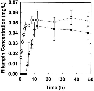
Penetration of 0.1 μg of rifampin per ml through membrane assemblies with (filled symbols) and without (open symbols) S. epidermidis colony biofilms. Error bars indicate the standard errors of the means.
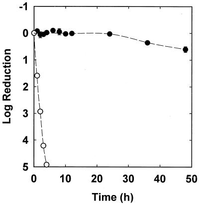
Comparison of susceptibilities of planktonic S. epidermidis (open symbols) and S. epidermidis in a 48-h colony biofilm (filled symbols) exposed to 0.1 μg of rifampin per ml. Error bars indicate the standard errors of the means.
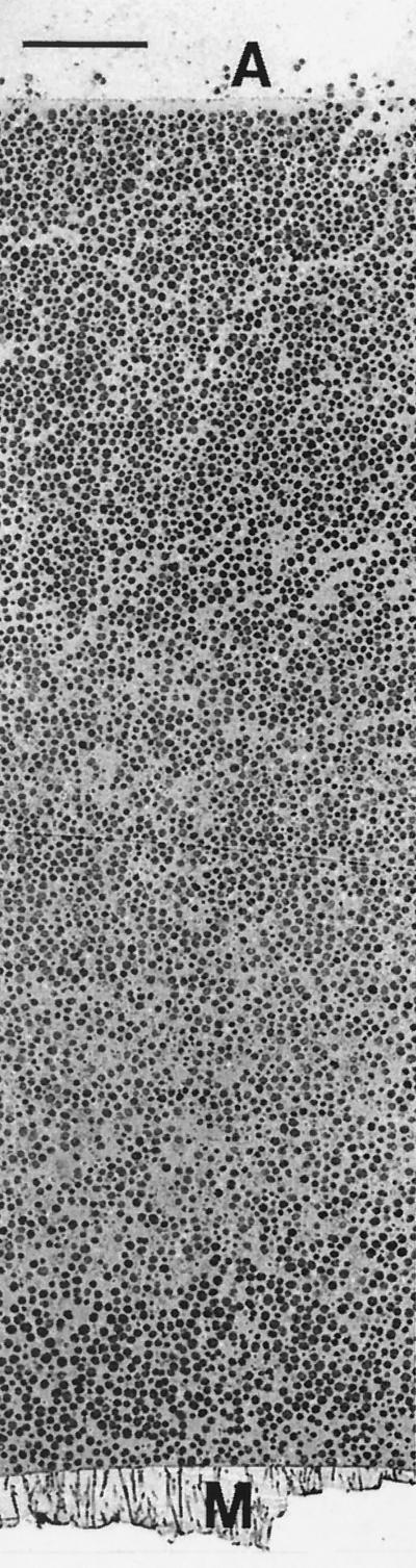
Transmission electron micrograph of the untreated, time zero control S. epidermidis colony biofilm. The membrane (M) is at the bottom, and the air interface (A) is at the top. Scale bar, 10 μm.
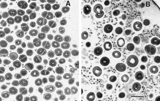
Transmission electron micrographs of S. epidermidis colony biofilms near the air interface. (A) Untreated 48-h control; (B) 48-h treatment with 0.1 μg of rifampin per ml. The air interface is located in the upper right-hand corners in both panels. Both images were taken at the same magnification. Scale bar, 1 μm.
References
-
- Andreoli-Pinto, T. J., and K. U. Graziano. 1999. Important aspects of the colonization of central venous catheter. Boll. Chim. Farm. 138:19-23. - PubMed
-
- Brown, M. R. W., D. G. Allison, and P. Gilbert. 1988. Resistance of bacterial biofilms to antibiotics: a growth-rate related effect? J. Antimicrob. Chemother. 22:777-783. - PubMed
-
- Cacoub, P., P. Leprince, P. Nataf, P. Hausfater, R. Dorent, B. Wechsler, V. Bors, A. Pavie, J. C. Peitte, and I. Gandjbakhch. 1998. Pacemaker infective endocarditis. Am. J. Cardiol. 82:480-484. - PubMed
-
- Costerton, J. W., P. S. Stewart, and E. P. Greenberg. 1999. Bacterial biofilms: a common cause of persistent infections. Science 284:1318-1322. - PubMed
MeSH terms
Substances
LinkOut - more resources
Full Text Sources
Other Literature Sources
Molecular Biology Databases
