. 2002 Feb 15;22(4):1208-17.
doi: 10.1523/JNEUROSCI.22-04-01208.2002.

Distribution and pharmacology of alpha 6-containing nicotinic acetylcholine receptors analyzed with mutant mice

Affiliations

Distribution and pharmacology of alpha 6-containing nicotinic acetylcholine receptors analyzed with mutant mice

Nicolas Champtiaux et al. J Neurosci. .

Abstract

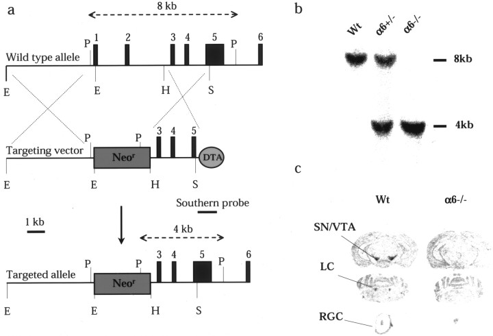
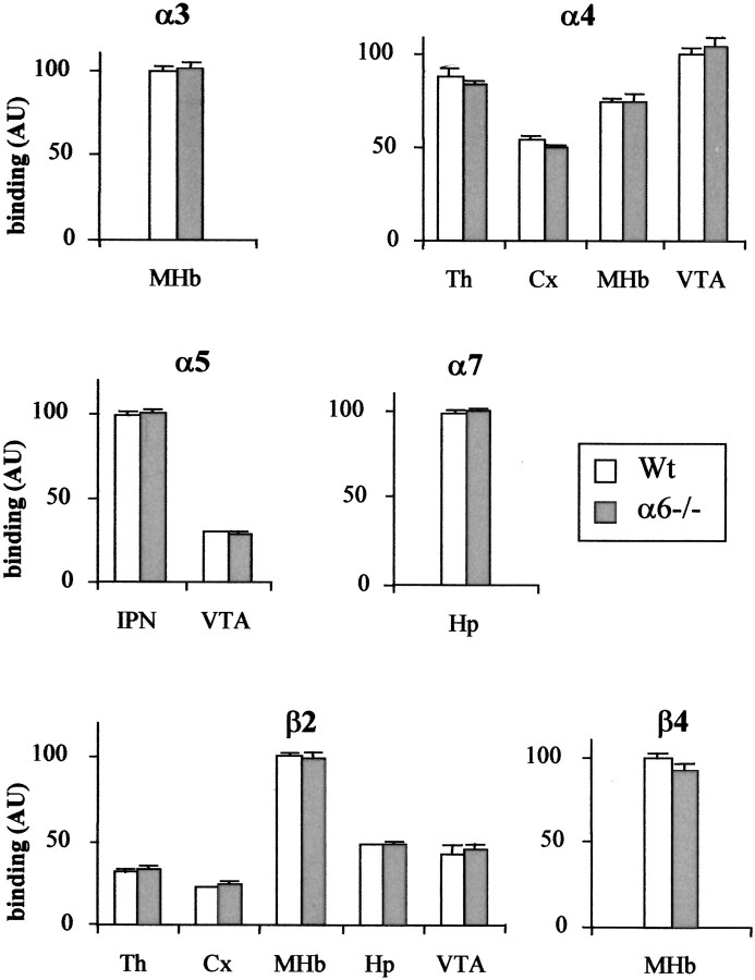
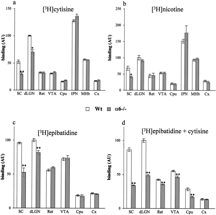
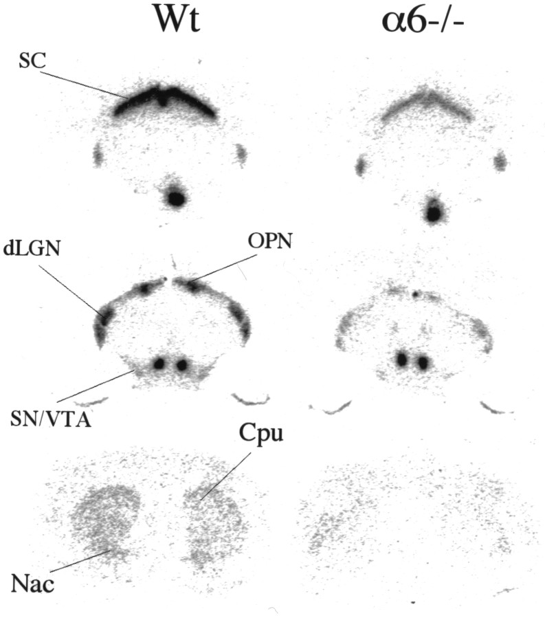
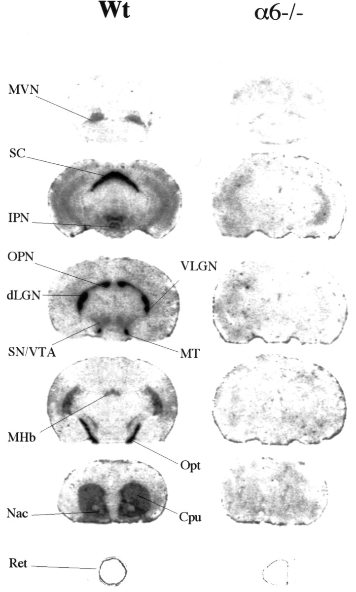
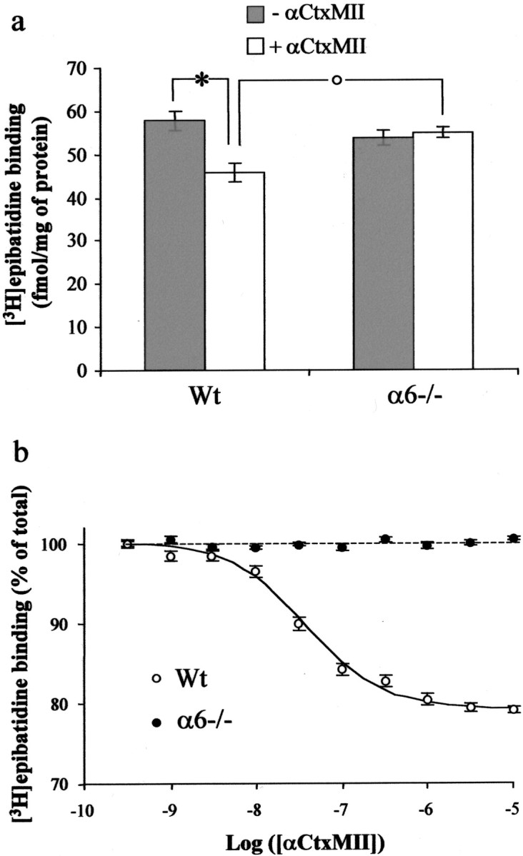
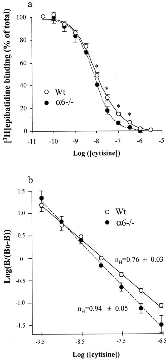
The alpha6 subunit of the nicotinic acetylcholine receptor (nAChR) is expressed at very high levels in dopaminergic (DA) neurons. However, because of the lack of pharmacological tools selective for alpha6-containing nAChRs, the role of this subunit in the etiology of nicotine addiction remains unknown. To provide new tools to investigate this issue, we generated an alpha6 nAChR knock-out mouse. Homozygous null mutants (alpha6-/-) did not exhibit any gross neurological or behavioral deficits. A careful anatomic and molecular examination of alpha6-/- mouse brains demonstrated the absence of developmental alterations in these animals, especially in the visual and dopaminergic pathways, where the alpha6 subunit is normally expressed at the highest levels. On the other hand, receptor autoradiography revealed a decrease in [3H]nicotine, [3H]epibatidine, and [3H]cytisine high-affinity binding in the terminal fields of retinal ganglion cells of alpha6-/- animals, whereas high-affinity [125I]alpha-conotoxinMII (alphaCtxMII) binding completely disappeared in the brain. Moreover, inhibition of [3H]epibatidine binding on striatal membranes, using unlabeled alphaCtxMII or cytisine, revealed the absence of alphaCtxMII-sensitive and cytisine-resistant [3H]epibatidine binding sites in alpha6-/- mice, although the total amount of binding was unchanged. Because alphaCtxMII, a toxin formerly thought to be specific for alpha3beta2-containing nAChRs, is known to partially inhibit nicotine-induced dopamine release, these results support the conclusion that alpha6 rather than alpha3 is the partner of beta2 in the nicotinic modulation of DA neurons. They further show that alpha6-/- mice might be useful tools to understand the mechanisms of nicotine addiction, although some developmental compensation might occur in these mice.


Figures

Fig. 1.
Fig. 1.
Gene targeting of the neuronal nAChR α6 subunit. a, Construction of the targeting vector. The homologous recombination event generated a 4 kb genomic deletion that removes exons 1 and 2. The targeting vector was designed to obtain a replacement mutation and contains a neomycin resistance gene (Neor) as a positive selection marker and the diphtheria toxin gene (DTA) as a negative selection marker. The expected wild-type and mutant restriction fragments after PstI digest and hybridization with the Southern blot probe are shown as dotted lines. Restriction enzymes are as follows: E,EcoRI; H, HindIII;P, PstI; S,SalI. b, Southern blot analysis of wild-type (Wt), heterozygous (α6+/−), and homozygous null mutant mice (α6−/−). Genomic tail DNA was digested withPstI and hybridized with anSalI–PstI probe, external to the targeting vector. The 4 kb band corresponds to the mutated allele.c, Detection of α6 mRNA in the brain of Wt and α6−/− animals, by in situ hybridization with an oligonucleotide located in the sixth exon of the α6 gene. In Wt animals, α6 mRNA is detected in SN–VTA neurons, LC and RGC. No signal above background levels is detected in α6−/− animals.
Fig. 2.
Fig. 2.
Semiquantitative analysis of α3, α4, α5, α7, β2, and β4 nAChR subunit mRNAs in Wt and Mt mice usingin situ hybridization. For each subunit, optical density value of the signal was measured and corrected for background and film linearity. This value was then normalized to 100 in the region where the signal was maximal. Results are expressed as means ± SEM of at least three animals. Statistical analysis was performed using an unpaired Student's t test (p < 0.05). Regions quantified were medial habenula (MHb), interpeduncular nucleus (IPN), thalamus (Th), cortex (Cx), VTA, and hippocampus (Hp). Note the absence of significant differences in the relative abundance of the mRNA of these subunits, in any brain region.
Fig. 3.
Fig. 3.
Quantitative autoradiography for nicotinic agonists in the mouse brain. Quantitative analysis of [3H]cytisine (a), [3H]nicotine (b), [3H]epibatidine (c), and [3H]epibatidine in the presence of 50 nm unlabeled cytisine (d), in Wt and α6−/− mice. The results are expressed as the mean specific optical density ± SEM, normalized to 100 in the dLGN of Wt animals. AU, Arbitrary unit. Statistical analysis was performed using an unpaired Student's t test (*p < 0.05; **p < 0.01) versus Wt controls. Regions quantified were SC, dLGN, retina (Ret), VTA, Cpu, IPN, MHb, and Cx.
Fig. 4.
Fig. 4.
Autoradiograms of cytisine-resistant [3H]epibatidine binding in the mouse brain. Sections were incubated with 400 pm[3H]epibatidine in the presence of 50 nm unlabeled cytisine. See Figure 3 for quantification.
Fig. 5.
Fig. 5.
Autoradiograms of [125I]αCtxMII binding in the mouse brain. Sections were incubated with 0.5 nm[125I]αCtxMII, as described in Materials and Methods. Residual signal seen on the eye section of α6−/− animals corresponds to nonspecific binding to pigmented epithelium. MT, Medial terminal nucleus of the accessory tract; MVN, medial vestibular nucleus; VLGN, ventrolateral geniculate nucleus.
Fig. 6.
Fig. 6.
Displacement of [3H]epibatidine binding by αCtxMII in mouse striatal membranes. a, Comparison of total (gray bars) and αCtxMII-resistant (10 μm, white bars) [3H]epibatidine binding sites in Wt and α6−/− animals. Each point represents the mean ± SEM of six separate determinations, expressed in femtomoles per milligram of protein. Statistical analysis was performed using an unpaired Student'st test. A statistically significant fraction (21%; *p < 0.01) of [3H]epibatidine binding sites is sensitive to αCtxMII displacement in Wt but not in α6−/− animals. αCtxMII-resistant [3H]epibatidine binding sites are significantly (°p < 0.01) upregulated in α6−/− animals compared with their Wt controls, whereas total [3H]epibatidine binding is unaffected by the genotype. b, Inhibition curve of [3H]epibatidine (500 pm) binding by αCtxMII (30 pm to 3 μm) in striatal membrane preparations of α6−/− (filled symbols) and Wt (open symbols) animals. Nonspecific binding, determined in the presence of 500 nmunlabeled epibatidine, was subtracted from each measurement. Results were then expressed as a percentage of specific binding in the absence of αCtxMII. Each value is the mean ± SEM of six separate determinations. See also Table 3.
Fig. 7.
Fig. 7.
Displacement of [3H]epibatidine binding by cytisine in mouse striatal membranes. a, Inhibition curve of [3H]epibatidine binding by cytisine: membrane samples were incubated with 500 pm[3H]epibatidine in the presence of (30 pm to 3 μm) unlabeled cytisine. Nonspecific binding, determined in the presence of 500 nmunlabeled epibatidine, was subtracted from each measurement. Results were then expressed as a percentage of specific binding in the absence of cytisine. Each point represents the mean ± SEM of five separate determinations. Data obtained from α6−/− (filled symbols) or Wt (open symbols) striatal membranes preparations were fitted to a one- (dotted line) or two-site inhibition model (see Material and Methods), respectively, depending on the value of the Hill number (see below). Statistical analysis was performed using an unpaired Student's t test (*p < 0.05).b, Hill plot representation. Log(B/(BoB)) is plotted as a function of log([cytisine]), whereB is the amount of specific [3H]epibatidine binding andBo is the specific binding in the absence of cytisine. In these conditions, the slope of the curve represents the Hill number, nH. In Wt animals,nH is significantly <1 (p < 0.05), suggesting the existence of an heterogeneous population of binding sites. Conversely, in α6−/− animals, nH = 0.94 ± 0.05, which suggests a homogeneous population of binding sites. See also Table4.

References

    1. Bansal A, Singer JH, Hwang BJ, Xu W, Beaudet A, Feller MB. Mice lacking specific nicotinic acetylcholine receptor subunits exhibit dramatically altered spontaneous activity patterns and reveal a limited role for retinal waves in forming ON and OFF circuits in the inner retina. J Neurosci. 2000;20:7672–7681. - PMC - PubMed
    1. Boye SM, Grant RJ, Clarke PB. Disruption of dopaminergic neurotransmission in nucleus accumbens core inhibits the locomotor stimulant effects of nicotine and d-amphetamine in rats. Neuropharmacology. 2001;40:792–805. - PubMed
    1. Cartier GE, Yoshikami D, Gray WR, Luo S, Olivera BM, McIntosh JM. A new alpha-conotoxin which targets alpha3beta2 nicotinic acetylcholine receptors. J Biol Chem. 1996;271:7522–7528. - PubMed
    1. Charpantier E, Barneoud P, Moser P, Besnard F, Sgard F. Nicotinic acetylcholine subunit mRNA expression in dopaminergic neurons of the rat substantia nigra and ventral tegmental area. NeuroReport. 1998;9:3097–3101. - PubMed
    1. Corrigall WA, Coen KM, Adamson KL. Self-administered nicotine activates the mesolimbic dopamine system through the ventral tegmental area. Brain Res. 1994;653:278–284. - PubMed

Publication types

MeSH terms