. 2002 Feb 15;22(4):1303-15.
doi: 10.1523/JNEUROSCI.22-04-01303.2002.

Small proline-rich repeat protein 1A is expressed by axotomized neurons and promotes axonal outgrowth

Affiliations

Small proline-rich repeat protein 1A is expressed by axotomized neurons and promotes axonal outgrowth

Iris E Bonilla et al. J Neurosci. .

Abstract

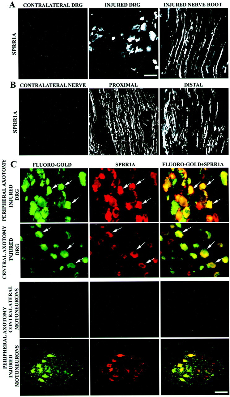
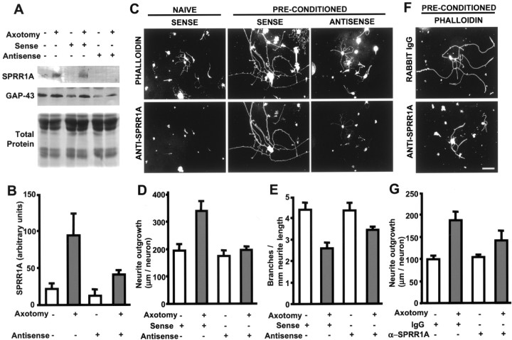
The ability of neurons to regenerate an axon after injury is determined by both the surrounding environment and factors intrinsic to the damaged neuron. We have used cDNA microarrays to survey those genes induced during successful sciatic nerve regeneration. The small proline-rich repeat protein 1A (SPRR1A) is not detectable in uninjured neurons but is induced by >60-fold after peripheral axonal damage. The protein is localized to injured neurons and axons. sprr1a is one of a group of epithelial differentiation genes, including s100c and p21/waf, that are coinduced in neurons by axotomy. Overexpressed SPRR1A colocalizes with F-actin in membrane ruffles and augments axonal outgrowth on a range of substrates. In axotomized sensory neurons, reduction of SPRR1A function restricts axonal outgrowth. Neuronal SPRR1A may be a significant contributor to successful nerve regeneration.


Figures

Fig. 1.
Fig. 1.
SPRR1A is induced in DRGs by axotomy.A, Northern blot analysis reveals that actin mRNA levels remain constant in lumbar DRGs 1 week after sciatic nerve lesion (LES) compared with control (CON) levels. DRGs dramatically upregulate both sprr1a andgap-43 mRNA after sciatic nerve transection. Migration of 28S and 18S ribosomal RNA is shown at left.B, Immunoblots for SPRR1A protein demonstrate upregulation in adult L3–L5 DRGs (1X, 10 μg total protein; 3X, 30 μg total protein) after transection of the ipsilateral sciatic nerve (LES) compared with unlesioned (CON) samples. SPRR1A is not detected in uninjured E15 DRG samples. There is a small but detectable increase in SPRR1A protein after thoracic SCI seen only in the 3X samples. Molecular weight markers are shown at left.C, DRGs contralateral to sciatic nerve axotomy do not express detectable SPRR1A by immunoblot (CONTROL,top). Nerve transection induces SPRR1A upregulation within 4 d after injury (middle). SPRR1A protein levels peak at 7–14 d, with a reduction 1 month after the injury. Nerve crush induces a similar SPRR1A expression level at 7 d, but levels decrease to baseline by 1 month (bottom). The number of days between nerve transection and animal death is shown at the top. Concentrations of SPRR1A protein in the DRG homogenates are reported at the bottom of each lane in nanograms of SPRR1A per microgram of total protein.
Fig. 2.
Fig. 2.
SPRR1A is expressed in regenerating sensory and motor neurons. A, SPRR1A immunofluorescence demonstrates protein in the cell bodies and axons of DRG neurons 1 week after peripheral axotomy (injured). No SPRR1A immunoreactivity can be found in adult control DRGs (contralateral). Scale bar, 50 μm.B, SPRR1A protein is distributed throughout DRG regenerating axons, as revealed by SPRR1A immunoreactivity of sciatic nerve 1 week after a crush injury. SPRR1A-positive axons were found up to 20 mm distal from the crush site. The protein is absent in the contralateral (uninjured) nerve. Scale bar, 100 μm (fromA). C, Intense SPRR1A protein immunoreactivity colocalizes with Fluoro-Gold retrogradely labeled sensory and motor neurons ipsilateral to a sciatic nerve transection (peripheral axotomy, arrows). SPRR1A is slightly elevated in sensory neurons 1 week after thoracic SCI (central axotomy). SPRR1A is absent from the ventral horn contralateral to a sciatic nerve transection. Scale bar: first andsecond rows, 50 μm; third andfourth rows, 100 μm.
Fig. 3.
Fig. 3.
S100C and p21/WAF1 are induced in regenerating sensory neurons by axotomy. A, Northern blot analysis ofs100c mRNA levels in lumbar DRGs from animals of different ages is shown. Adult DRG samples were analyzed contralateral (CON) or ipsilateral (LES) to a sciatic nerve transection 1 week before death. A clear upregulation of the transcript after axotomy is evident. p21/waf1 mRNA levels are regulated in a similar manner in the lower panel. Two micrograms of total RNA were loaded in each lane.B, S100C immunoblots demonstrate protein levels contralateral (CONTROL) or ipsilateral (TRANSECTION) to a sciatic nerve transection at the indicated times after lesion. Quantification of the relative levels of S100C protein in the axotomized DRG samples is reported at thebottom. C, In situhybridization demonstrates s100c andp21/waf1 mRNA expression in lumbar DRGs contralateral or ipsilateral (injured) to sciatic nerve transection 1 week before death.D, S100C immunostaining of DRG sections that were retrogradely labeled with Fluoro-Gold demonstrates that the protein is induced selectively in neurons that underwent transection 1 week earlier at the midthigh (arrows). Scale bar, 50 μm.
Fig. 4.
Fig. 4.
SPRR1A colocalization with F-actin and S100C in COS-7 cells and neuronal growth cones. A, COS-7 cells were transfected with pCDNA3.1-SPRR1A-Myc. SPRR1A immunoreactivity colocalizes with F-actin-rich structures, predominantly at dorsal and leading edge ruffles (arrowheads). SPRR1A is absent from actin-rich stress fibers (arrows). A–C, The left three panels show double labeling of one cell, and the extreme right panels show a different cell.B, COS-7 cells were transfected with pCDNA3.1-S100C-Myc-His. S100C immunoreactivity colocalizes with F-actin (ruffles, arrowheads). C, pCDNA3.1-SPRR1A-Myc and pDNA3.1-S100C-Myc-His were cotransfected into COS-7 cells. SPRR1A and S100C immunoreactivity codistributed at leading edge and dorsal ruffles (arrowheads). S100C was detected by staining with monoclonal anti-His antibodies. D, The distribution of SPRR1A and F-actin in a pDNA3.1-SPRR1A-Myc-transfected COS-7 cell is examined in three dimensions. Top, Vertical (z-axis) cross section at the level of theblue arrows for the lower three panels. The numbers on the lower three panels refer to the distance above the substrate. Note the predominant distribution of SPRR1A to dorsal and leading edge ruffles (arrowheads). A dorsal ruffle is shown to protrude into the upper region of the cell.E, Chick E7 DRG cultures were infected with HSV-SPRR1A and examined for SPRR1A and F-actin distribution 24 hr later by confocal microscopy. In axonal growth cones (left three panels show one growth cone, and the next panelshows a second growth cone) and fibroblasts (right panel), note the similar distribution of SPRR1A and F-actin in linear aggregates and ruffles (arrowheads). Other F-actin-rich structures (arrows), such as stress fibers, are not enriched in SPRR1A. Scale bar, 50 μm.
Fig. 5.
Fig. 5.
SPRR1A promotes axonal outgrowth in embryonic neurons. A, Endogenous SPRR1A protein immunoreactivity is not detected in chick E7 and adult mouse DRGs cultured for 1 DIV but is present after 5 DIV by immunofluorescence. Scale bar, 100 μm. B, Overexpression of SPRR1A and S100C protein in chick E7 DRGs via recombinant HSV infection increases axonal growth compared with HSV-EGFP-infected cells. EGFP-, SPRR1A-, and S100C-expressing neurons are identified by EGFP fluorescence, SPRR1A immunoreactivity, and S100C immunoreactivity, respectively.C, Neurite outgrowth was determined in neurons expressing EGFP, Nogo-66 receptor (NgR, as a control), or SPRR1A via recombinant HSV infection. A significant (p ≤ 0.05, Student's two-tailedt test) increase in outgrowth is observed in HSV-SPRR1A-infected cultures 24 hr after plating. Data are means ± SEM from five experiments. D, DRG neurons infected with HSV-EGFP or HSV-SPRR1A were cultured on the indicated concentrations of laminin. Mean neurite outgrowth per infected neuron is reported (± SEM). E, A significant (p ≤ 0.05, Student's two-tailedt test) increase in neurite outgrowth (± SEM) is observed in neurons triturated with purified SPRR1A. Control triturations have no effect on outgrowth. SPRR1A protein added to the culture medium without trituration does not alter outgrowth.F, Neurite outgrowth was measured for neurons overexpressing EGFP and S100C via recombinant HSV infection. A significant (p ≤ 0.05, Student's two-tailed t test) increase in outgrowth is observed for the HSV-S100C-infected neurons 24 hr after plating.
Fig. 6.
Fig. 6.
SPRR1A promotes axonal outgrowth in adult neurons and on inhibitory substrates. A, Phalloidin staining of adult mouse DRGs after 1 DIV illustrates the different modes of growth that characterize naive and preconditioned neurons. Preconditioned DRGs were removed and plated 4 d after sciatic nerve axotomy. Whereas naive DRGs extend short and highly branched neurites, preconditioned DRGs grow neurites that are elongated and sparsely branched.B, Overexpression of SPRR1A protein in adult mouse DRGs via recombinant HSV infection increases axonal growth and decreases branching compared with HSV-EGFP-infected cells. EGFP- and SPRR1A-expressing neurons are identified by EGFP fluorescence and SPRR1A immunoreactivity, respectively. Scale bar, 100 μm.C, Neurite outgrowth was determined in naive and preconditioned adult mouse DRG neurons (phalloidin staining) and in neurons expressing EGFP (as a control) or SPRR1A via recombinant HSV infection. A significant (p ≤ 0.05, Student's two-tailed t test) increase in outgrowth is observed in HSV-SPRR1A-infected cultures 24 hr after plating. Data are means ± SEM from three experiments. D, Axonal branching was calculated for naive and preconditioned neurons and for HSV-EGFP- and HSV-SPRR1A-infected DRGs. We confirmed that preconditioning and HSV-SPRR1A infection significantly (p ≤ 0.05, Student's two-tailedt test) decrease branching compared with naive neurons, and a parallel decrease was observed in HSV-SPRR1A-infected DRGs. Data are means ± SEM from three experiments. E, Neurite outgrowth was measured for HSV-EGFP- and HSV-SPRR1A-infected E13 DRG neurons plated on laminin or GST-Nogo-66 as the substrate. HSV-SPRR1A-infected neurons exhibit increased outgrowth relative to HSV-EGFP-infected neurons (p ≤ 0.05, Student's two-tailed t test) when plated on laminin (10 μg/ml) or on Nogo (34 ng/mm2). Data are means ± SEM from five experiments. F, Neurite outgrowth was measured for HSV-EGFP- and HSV-SPRR1A-infected E13 DRG neurons plated on laminin or bovine CNS myelin as the substrate. HSV-SPRR1A-infected neurons show increased outgrowth compared with HSV-EGFP-infected neurons (p ≤ 0.05, Student's two-tailed t test) when plated on laminin (10 μg/ml) or on CNS myelin (45 ng/mm2). Data are means ± SEM from five experiments.
Fig. 7.
Fig. 7.
SPRR1A loss of function decreases axonal regeneration in adult DRGs. A, Immunoblots for SPRR1A and GAP-43 protein from adult mouse DRG neurons are illustrated. Naive or preconditioned neurons were cultured with sense or antisense oligonucleotides, as indicated. A decrease in SPRR1A immunoreactivity but not in GAP-43 levels is observed after treatment with antisense oligonucleotide. Coomassie brilliant blue staining reveals that total protein levels are equal in all samples. B, The SPRR1A signal from immunoblots of control and antisense-treated DRG cultures as in A is quantified. The level of SPRR1A protein decreases to nearly basal levels in neurons treated with antisense oligonucleotide. Data are means ± SEM from three experiments.C, Adult mouse DRG neurons in the naive or preconditioned state were cultured with sense or antisense oligonucleotides. Each vertical pair of panels shows the same field double-labeled with phalloidin to reveal F-actin or with anti-SPRR1A. Naive neurons treated with sense oligonucleotides extend short, branched processes without SPRR1A protein, whereas preconditioned neurons display elongated, less branched axons expressing SPRR1A protein. Preconditioned neurons treated with antisense oligonucleotides exhibit little anti-SPRR1A staining and morphological features similar to those of naive neurons. D, Neurite outgrowth was measured for naive and preconditioned neurons treated with either sense or antisense oligonucleotides. A significant (p ≤ 0.05, Student's two-tailedt test) decrease in axonal length is observed for antisense oligonucleotide-treated preconditioned neurons compared with sense-treated preconditioned neurons. Data are means ± SEM from three experiments. E, Axonal branching was determined for naive and preconditioned adult mouse DRG neurons treated with sense or antisense oligonucleotides. For naive neurons, oligonucleotide treatment did not alter branching. For preconditioned neurons, antisense oligonucleotide treatment increased branching compared with sense-treated neurons (p ≤ 0.05, Student's two-tailed t test). Data are means ± SEM from three experiments. F, Neurite outgrowth was examined in preconditioned adult neuronal cultures triturated in the presence of rabbit IgG or α-SPRR1A. Axonal length was decreased after antibody-mediated blockade of SPRR1A protein compared with rabbit IgG. Scale bar, 100 μm. G, Axonal growth was measured in naive and preconditioned neurons triturated with control antibody, rabbit IgG (0.2 mg/ml), or affinity-purified SPRR1A antibody (0.2 mg/ml). Neurite length was decreased in α-SPRR1A-triturated preconditioned neurons relative to IgG-treated preconditioned neurons (p ≤ 0.05, Student's two-tailedt test). No change in neurite outgrowth was observed in naive neurons triturated with α-SPRR1A. Data are means ± SEM from three experiments.

References

    1. Allen BG, Durussel I, Walsh MP, Cox JA. Characterization of the Ca2+-binding properties of calgizzarin (S100C) isolated from chicken gizzard smooth muscle. Biochem Cell Biol. 1996;74:687–694. - PubMed
    1. Bomze HM, Bulsara KR, Iskandar BJ, Caroni P, Skene JH. Spinal axon regeneration evoked by replacing two growth cone proteins in adult neurons. Nat Neurosci. 2001;4:38–43. - PubMed
    1. Chong MS, Woolf CJ, Haque NS, Anderson PN. Axonal regeneration from injured dorsal roots into the spinal cord of adult rats. J Comp Neurol. 1999;410:42–54. - PubMed
    1. Cox LS. Multiple pathways control cell growth and transformation: overlapping and independent activities of p53 and p21Cip1/WAF1/Sdi1. J Pathol. 1997;183:134–140. - PubMed
    1. Dickson BJ. Rho GTPases in growth cone guidance. Curr Opin Neurobiol. 2001;11:103–110. - PubMed

Publication types

MeSH terms