. 2002 Feb 15;22(4):1316-27.
doi: 10.1523/JNEUROSCI.22-04-01316.2002.

Brain-derived neurotrophic factor modulates cerebellar plasticity and synaptic ultrastructure

Affiliations

Brain-derived neurotrophic factor modulates cerebellar plasticity and synaptic ultrastructure

Alexandre R Carter et al. J Neurosci. .

Abstract

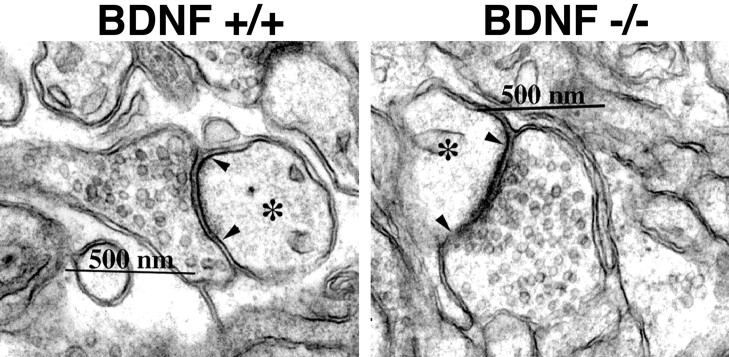
Neurotrophins are key regulators of neuronal survival and function. Here we show that TrkB, the receptor for brain-derived neurotrophic factor (BDNF), is located at parallel fiber to Purkinje cell (PF/PC) synapses of the cerebellum. To determine the effects of TrkB receptor activation on synapse formation and function, we examined the parallel fiber to Purkinje cell synapses of mice with a targeted deletion of the BDNF gene. Although Purkinje cell dendrites are abnormal in BDNF -/- mice, PF/PC synapses are still able to form. Immunohistochemical analysis of mutant animals revealed the formation of numerous PF/PC synapses with the appropriate apposition of presynaptic and postsynaptic proteins. These synapses are functional, and no differences were detected in the waveform of evoked EPSCs, the amplitude of spontaneous mini-EPSCs, or the response to prolonged 10 Hz stimulus trains. However, paired-pulse facilitation, a form of short-term plasticity, is significantly decreased in BDNF -/- mice. Detailed ultrastructural analysis of the presynaptic terminals demonstrated that this change in synaptic function is accompanied by an increase in the total number of synaptic vesicles in mutant mice and a decrease in the proportion of vesicles that are docked. These data suggest that BDNF regulates both the mechanisms that underlie short-term synaptic plasticity and the steady-state relationship between different vesicle pools within the terminal.


Figures

Fig. 1.
Fig. 1.
The BDNF receptor, TrkB, is located at PF/PC synapses. A, P15 cerebellum immunolabeled with anti-TrkB. TrkB staining is seen throughout the molecular layer (ML) and in Purkinje cell bodies (PC) and dendrites in BDNF +/+ mice. EGL, External granule cell layer; IGL, internal granule cell layer. B,C, Single optical section through the molecular layer of P15 wild-type cerebellum labeled with anti-calbindin before deconvolution (B) and after deconvolution (C). DF, Cerebellar molecular layer of P15 wild-type mouse double labeled with anti-calbindin (D, green) and anti-TrkB (E, red) antibodies. The merged image (F) shows numerous TrkB puncta juxtaposed to and sometimes overlapping with calbindin-positive Purkinje cell dendritic spines (arrowheads). GI, Cerebellar molecular layer of P15 wild-type mouse double labeled with anti-calbindin (G, green) and anti-GluRδ2 (GluR delta 2) (H,red). Merged image (I) shows GluRδ2-positive overlapping Purkinje cell dendrites and capping Purkinje cell dendritic spines (arrowheads).JL, Cerebellar molecular layer of P15 wild-type mouse double labeled with anti-SV2 (J,green) and anti-TrkB (K,red). Merged image (L) shows SV2-positive puncta juxtaposed to and partially overlapping with TrkB-positive puncta (arrowheads).DL were reconstructed from 5–10 adjacent optical sections. Scale bars: A, 20 μm;B, C, 10 μm;DI, 5 μm.;JL, 5 μm.
Fig. 2.
Fig. 2.
The development of Purkinje cell dendritic arbors in wild-type (A, C, D) and BDNF −/− mice (B, D,F). Parasagittal cerebellar sections from P8 (A, B), P15 (C,D), and P24 (E, F) mice were immunolabeled with anti-calbindin antibody. In the absence of BDNF, Purkinje cell dendrites are stunted at P8 and undergo significant development over the following 2 weeks, but remain less extensive than in WT mice. Scale bar, 20 μm.
Fig. 3.
Fig. 3.
Individual Purkinje cell dendrite morphology in the absence of BDNF. A, Modified Golgi stain resolves the morphology of an individual Purkinje cell dendritic arbor in a P15 wild-type mouse. Inset shows a higher magnification view of a dendritic segment studded with numerous spines (arrowheads). B, C, Two individual Purkinje cells from a P15 BDNF −/− mouse exhibit a significant decrease in the extent of the dendritic arbor.Insets show that dendrites nonetheless carry numerous spines (arrowheads). Scale bars: A–C, 20 μm; insets, 5 μm.
Fig. 4.
Fig. 4.
Synaptogenesis in the molecular layer of BDNF −/− cerebellum. A, P15 cerebellum immunolabeled with antibodies against calbindin (green) and the glutamate receptor subunit GluRδ2 (GluR delta 2) (red). Numerous calbindin-positive spines are capped by puncta of GluRδ2 staining characteristic of functional synapses.B, Individual presynaptic terminals in the molecular layer immunolabeled with antibodies against the presynaptic terminal proteins SV2 (green) and synapsin (red) resulting in identical staining patterns.C, Immunolabeling with antibodies against SV2 (green) and GluRδ2 (red) results in staining patterns where the puncta are juxtaposed with much less overlap, consistent with the association of presynaptic terminals with their postsynaptic targets. Images were reconstructed from 5–10 adjacent optical sections. Scale bars: A, 5 μm;B, C, 1 μm.
Fig. 5.
Fig. 5.
A, Representative consecutive mEPSC traces are shown from a wild-type (left) and a BDNF −/− (right) mouse. B, The normalized cumulative amplitude distribution for BDNF +/+ (n = 4; solid trace) and BDNF −/− (n = 4; dashed trace) are similar (p = 1.0 by Kolmogorov–Smirnov test).
Fig. 6.
Fig. 6.
Specific impairment of short-term synaptic plasticity in BDNF −/− mice. A, Evoked AMPA receptor-mediated EPSCs were elicited by stimulating parallel fibers and recording from an individual Purkinje cell in voltage clamp. The decay times of the evoked EPSCs are indistinguishable between WT and BDNF −/− mice. Traces are the averages of 5–10 trials.B, The mean percentage facilitation is plotted as a function of interstimulus intervals for BDNF −/− (▪;n = 6) and wild-type littermates (■;n = 6). BDNF −/− mice exhibit decreased paired-pulse facilitation at interstimulus intervals <200 msec (p < 0.05). C, The response of Purkinje cells to repetitive 10 Hz stimulation is illustrated by plotting the ratio of the amplitude of the response to the 10th and 50th pulses to the amplitude of the response to the first pulse. Although 10th/1st ratio is significantly decreased in BDNF −/− mice because of impaired PPF, there is no significant difference in the 50th/1st. This indicates that there is no difference in the response to prolonged 10 Hz stimulation in the absence of BDNF.
Fig. 7.
Fig. 7.
Electron micrographs of the PF/PC synapse in P15 WT and BDNF −/− mice. Individual synapses exhibit a dendritic spine (∗), postsynaptic density demarcated by arrowheads, and presynaptic terminal filled with numerous round, loosely packed vesicles in both wild-type and BDNF −/− animals.
Fig. 8.
Fig. 8.
Increased vesicle number in BDNF −/− synapses is restricted to vesicles located farthest from the active zone. The number of vesicles within consecutive 25 nm bins was plotted as a function of distance from the synaptic cleft. Within the first 100 nm from the synaptic cleft there is no difference in the number of vesicles between the wild-type mice (▪) and BDNF −/− mice (■) in either longitudinal sections (A) or cross sections (B). However, beyond 100 nm, BDNF −/− mice exhibit a significant increase in the number of vesicles compared with WT mice. * DD0.01by Kolmogorov–Smirnov.

References

    1. Atluri PP, Regehr WG. Delayed release of neurotransmitter from cerebellar granule cells. J Neurosci. 1998;18:8214–8227. - PMC - PubMed
    1. Berninger B, Schinder AF, Poo MM. Synaptic reliability correlates with reduced susceptibility to synaptic potentiation by brain-derived neurotrophic factor. Learn Mem. 1999;6:232–242. - PMC - PubMed
    1. Betz WJ, Angleson JK. The synaptic vesicle cycle. Annu Rev Physiol. 1998;60:347–363. - PubMed
    1. Bolshakov VY, Siegelbaum SA. Regulation of hippocampal transmitter release during development and long-term potentiation. Science. 1995;269:1730–1734. - PubMed
    1. Bridgman PC. Myosin Va movements in normal and dilute-lethal axons provide support for a dual filament motor complex. J Cell Biol. 1999;146:1045–1060. - PMC - PubMed

Publication types

MeSH terms

LinkOut - more resources