. 2002 Feb 15;539(Pt 1):3-14.
doi: 10.1113/jphysiol.2001.013303.

Regulation of the glutamine transporter SN1 by extracellular pH and intracellular sodium ions

Affiliations

Regulation of the glutamine transporter SN1 by extracellular pH and intracellular sodium ions

Angelika Bröer et al. J Physiol. .

Abstract

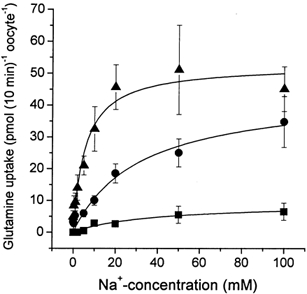
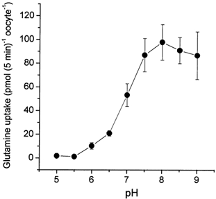
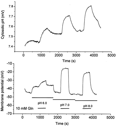
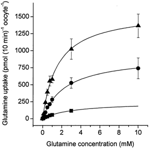
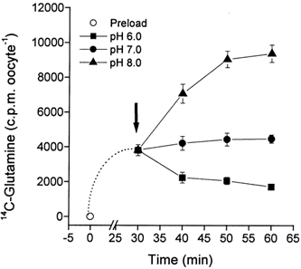
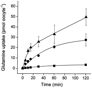
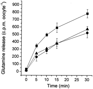
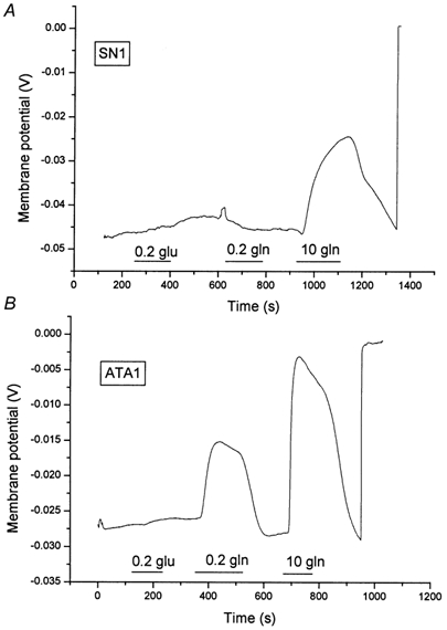
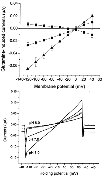
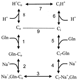
The glutamine transporter SN1 has recently been identified as one of the major glutamine transporters in hepatocytes and brain astrocytes. It appears to be the molecular correlate of system N amino acid transport. Two different transport mechanisms have been proposed for this transporter. These are an electroneutral mechanism, in which glutamine uptake is coupled to an exchange of 1Na+ and 1H+, or an electrogenic mechanism coupled to the exchange of 2Na+ against 1H+. This study was performed to solve these discrepancies and to investigate the reversibility of the transporter. When SN1 was expressed in Xenopus laevis oocytes, glutamine uptake was accompanied by a cotransport of 2-3 Na+ ions as determined by 22Na+ fluxes. However, at the same time a rapid release of intracellular Na+ was observed indicating an active exchange of Na+ ions. The driving force of the proton electrochemical gradient was equivalent to that of the sodium electrochemical gradient. Acidification of the extracellular medium caused the transporter to run in reverse and to release glutamine. Determination of accumulation ratios at different driving forces were in agreement with an electroneutral 1Na+-glutamine cotransport-1H+ antiport. Inward currents that were observed during glutamine uptake were much smaller than expected for a stoichiometric cotransport of charges. A slippage mode in the transporter mechanism and pH-regulated endogenous oocyte cation channels are likely to contribute to the observed currents.


Figures

Figure 1
Figure 1. Dependence of glutamine uptake via SN1 on the extracellular Na+ concentration at different pH values
Oocytes were injected with 20 ng SN1 cRNA or remained uninjected. After an incubation period of 4 days, uptake of labelled glutamine (100 μm) was determined over a period of 10 min in buffers of different NaCl concentration (NaCl replaced by NMDG-chloride) at pH 6.0 (filled squares), pH 7.0 (filled circles) and pH 8.0 (filled triangles). The transport activity of non-injected oocytes is already subtracted. The mean transport activity of 10 oocytes was determined for each datapoint.
Figure 2
Figure 2. Release of 22Na+ during glutamine uptake
Oocytes were injected with 20 ng SN1 cRNA or remained uninjected. A, after an incubation period of 6 days, 10 oocytes were first preloaded with 22Na+ (10 mmNaCl, 86 mm NMDG-Cl) at pH 7.4 in the presence of 10 mm glutamine. After 10 min oocytes were washed and the transport buffer was replaced by the same unlabelled buffer in the continued presence of 10 mm glutamine. Release of 22Na+ was followed by taking samples from the supernatant. In 10 control oocytes of the same batch the level of 22Na+ preloading was determined. The maximum releasable pool of 22Na+ is shown by the horizontal line in the graph. B, in a different experiment 10 oocytes were first preloaded with 22Na+ (10 mmNaCl, 86 mm NMDG-Cl) at pH 7.4 in the presence of 10 mm glutamine (preloading level 14600 ± 2500 c.p.m.). After 10 min oocytes were washed and the transport buffer was replaced by the same unlabelled buffer with or without addition of 10 mm glutamine. The intracellular Na+ that remained after 30 min of efflux in the oocytes was determined under both conditions. The difference in scale between experiments A and B resulted from the differing specific activity of the 22NaCl batches.
Figure 3
Figure 3. Glutamine transport via SN1 is pH dependent
Oocytes were injected with 20 ng SN1 cRNA or remained uninjected. After an incubation period of 4 days, uptake of labelled glutamine (100 μm) was determined over a period of 5 min. The transport activity of non-injected oocytes is already subtracted. The mean transport activity of 10 oocytes was determined for each pH value.
Figure 4
Figure 4. Uptake of glutamine via SN1 increases the cytosolic pH of oocytes
Oocytes were injected with 20 ng SN1 cRNA or remained uninjected. After an incubation period of 3 days, oocytes were superfused with glutamine (10 mm)-containing or glutamine-free solutions of different pH values. The cytosolic pH (upper panel) and the membrane potential (lower panel) were recorded with microelectrodes. Substrate superfusion periods are indicated by horizontal bars. Non-injected oocytes did not respond to superfusion of glutamine.
Figure 5
Figure 5. Determination of the glutamine Km at different pH values
Oocytes were injected with 20 ng SN1 cRNA or remained uninjected. After an incubation period of 4 days, uptake of labelled glutamine was determined over a period of 10 min. The glutamine concentration was varied between 0 and 10 mm in transport buffers titrated to pH 6.0 (filled squares), pH 7.0 (filled circles) and pH 8.0 (filled triangles). The transport activity of non-injected oocytes is already subtracted. The mean transport activity of 10 oocytes was determined for each datapoint. The 10 mm data point in the pH 6.0 set could not be evaluated due to the low specific activity.
Figure 6
Figure 6. Glutamine transport via SN1 reverses at acidic pH
Oocytes were injected with 20 ng SN1 cRNA or remained uninjected. After an incubation period of 3 days, oocytes were first preloaded with labelled glutamine (100 μm) at pH 7.4 (open circle). After 30 min preloading, the ‘pH switch buffer’ was added to the samples to adjust the final pH to 6.0 (filled squares), pH 7.0 (filled circles) or pH 8.0 (filled triangles). The ‘pH switch buffer’ differed in pH from the preloading buffer, but otherwise had an identical substrate concentration, specific activity and salt concentration.
Figure 7
Figure 7. Accumulation of glutamine at different extracellular pH values
Oocytes were injected with 20 ng SN1 cRNA or remained uninjected. After an incubation period of 4 days, uptake of labelled glutamine (10 μm) was determined in transport buffers adjusted to pH 6.0 (filled squares), pH 7.0 (filled circles) or pH 8.0 (filled triangles). Samples were taken at the indicated time points. The transport activity of non-injected oocytes is already subtracted. The mean transport activity of 10 oocytes was determined for each datapoint. The glutamine accumulation of non-injected oocytes is already subtracted.
Figure 8
Figure 8. Efflux of glutamine at different extracellular pH values
Oocytes were injected with 20 ng SN1 cRNA or remained uninjected. After an incubation period of 4 days, oocytes were injected with [14C]glutamine (final concentration 1 mm). Oocytes were washed with 4 ml ND96 buffer and then suspended in 1 ml transport buffer adjusted to pH 6.0 (filled squares), pH 7.0 (filled circles) or pH 8.0 (filled triangles). Samples were taken from the supernatant at the indicated time points. The mean efflux activity of four different experiments is shown in the graph. Efflux in non-injected oocytes was less than 10% of efflux observed in SN1 expressing oocytes.
Figure 9
Figure 9. Depolarization of the membrane potential during transport of glutamine via SN1 and ATA1
Oocytes were injected with 20 ng SN1 cRNA (A) or 20 ng ATA1 cRNA (B). After an incubation period of 4 days, oocytes were superfused with glutamine (0.2 mm and 10 mm) and glutamate containing solutions (0.2 mm). The membrane potential was recorded with microelectrodes. At the end of each recording microelectrodes were pulled out of the oocyte to record the bath potential. Superfusion periods are indicated by the horizontal bars. Non-injected oocytes did not respond to glutamate or glutamine.
Figure 10
Figure 10. Voltage dependence of glutamine-induced currents at different pH values
Oocytes were injected with 20 ng SN1 cRNA. After an expression period of 3 days oocytes were superfused with ND96 containing 10 mm glutamine or control solution (ND96) adjusted to pH 6.0 (filled squares), pH 7.0 (filled circles) and pH 8.0 (filled triangles). Once currents (in the absence or presence of substrate) remained stable, voltage ramps were run clamping the membrane potential from −120 mV to + 60 mV. The graph (upper panel) depicts the difference of the elicited currents in the presence and absence of substrate. The lower panel shows recordings from a representative oocyte. The thick lines show traces recorded in the presence of 10 mm glutamine (pH indicated to left), the thin traces were recorded in the absence of substrate. The holding potential during recording is given on the abscissa.
Figure 11
Figure 11. A kinetic model of glutamine transport via SN1
Experimental observations can be explained with an ordered binding model in which glutamine binds before Na+, allowing Na+ exchange (steps 2, 3 and 4). Slippage of the unloaded transporter (dotted line), creates an electrogenic transport mode that is similar to system A (steps 1, 2, 3, 4, 5 and 9). The normal transport cycle includes steps 1–8.

Similar articles

Cited by

References

    1. Albers A, Bröer A, Wagner CA, Setiawan I, Lang P, Kranz EU, Lang F, Bröer S. Na+ transport by the neural glutamine transporter ATA1. Pflügers Archiv. 2001;443:92–101. - PubMed
    1. Bröer S, Brookes N. Transfer of glutamine between astrocytes and neurons. Journal of Neurochemistry. 2001;77:705–719. - PubMed
    1. Bröer S, Schneider HP, Broer A, Rahman B, Hamprecht B, Deitmer JW. Characterization of the monocarboxylate transporter 1 expressed in Xenopus laevis oocytes by changes in cytosolic pH. Biochemical Journal. 1998;333:167–174. - PMC - PubMed
    1. Bröer A, Wagner C, Lang F, Bröer S. Neutral amino acid transporter ASCT2 displays substrate-induced Na+ exchange and a substrate-gated anion conductance. Biochemical Journal. 2000a;346:705–710. - PMC - PubMed
    1. Bröer A, Wagner C, Lang F, Bröer S. The heterodimeric amino acid transporter 4F2hc/y+LAT2 mediates arginine efflux in exchange with glutamine. Biochemical Journal. 2000b;349:787–795. - PMC - PubMed

Publication types

LinkOut - more resources