. 2002 Feb 15;539(Pt 1):77-91.
doi: 10.1113/jphysiol.2001.013258.

Nitric oxide inhibits capacitative Ca2+ entry and enhances endoplasmic reticulum Ca2+ uptake in bovine vascular endothelial cells

Affiliations

Nitric oxide inhibits capacitative Ca2+ entry and enhances endoplasmic reticulum Ca2+ uptake in bovine vascular endothelial cells

Elena N Dedkova et al. J Physiol. .

Abstract

In vascular endothelial cells, elevation of cytosolic free calcium concentration ([Ca2+]i) causes activation of nitric oxide synthase (NOS) and release of nitric oxide (NO). The goal of the study was to characterize the interplay between [Ca2+]i and NO production in this cell type. Simultaneous measurements of [Ca2+]i and intracellular NO concentration ([NO]i) in cultured bovine vascular endothelial cells (CPAE cell line) with the fluorescent indicators fura-2 and DAF-2, respectively, revealed that Ca2+ influx following agonist-induced intracellular Ca2+ store depletion (capacitative Ca2+ entry, CCE) represents the preferential Ca2+ source for the activation of the Ca2+-calmodulin-dependent endothelial NOS (eNOS). Exposure to the NO donor sodium nitroprusside (SNP) showed that high NO levels suppressed CCE and had an inhibitory effect on Ca2+ extrusion by the plasmalemmal Ca2+-ATPase. This inhibitory effect on CCE was mimicked by the membrane-permeant cGMP analogue 8-bromo-cGMP, but was reversed by the NO scavenger haemoglobin and prevented by the inhibitor of the NO-sensitive guanylate cyclase ODQ. Brief exposure to SNP reduced the peak of ATP-induced Ca2+ release from the endoplasmic reticulum (ER) and accelerated Ca2+ reuptake into the ER. Prolonged incubation with SNP resulted in enhanced Ca2+ loading of the ER, as revealed by direct measurements of store content with the ER-entrapped low-affinity Ca2+ indicator mag-fura-2. The results suggest that in vascular endothelial cells, NO synthesis is under autoregulatory control that involves NO-dependent [Ca2+]i regulation. Via cGMP-dependent inhibition of CCE and acceleration of Ca2+ sequestration into the ER, NO can lower [Ca2+]i and therefore exert an autoregulatory negative feedback on its own Ca2+-dependent synthesis.


Figures
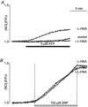
Figure 1
Figure 1. Effect of NOS inhibition on the ATP- and SNP-induced [NO]i elevations
A, ATP (5 μm) induced an increase of [NO]i that was completely abolished when cells were preincubated with the NOS inhibitor l-NNA (100 μm). The control trace represents basal [NO]i levels in the absence of ATP. B, effect of exposure to SNP on [NO]i in the presence and absence of l-NNA. Cells were pretreated with l-NNA for > 40 min before exposure to SNP. DAF-2 fluorescence intensities in each experiment were normalized to the level of fluorescence recorded prior to stimulation.
Figure 2
Figure 2. Simultaneous recordings of [Ca2+]i and [NO]i in CPAE cells
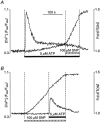
A, ATP stimulation (5 μm) induced a biphasic [Ca2+]i increase and NO production that became more prominent during the plateau phase of the [Ca2+]i transient. Subsequent SNP (100 μm) exposure significantly increased [NO]i level and suppressed the sustained [Ca2+]i plateau phase. B, high NO levels, achieved by SNP application 120 s before ATP stimulation, significantly decreased the ATP-induced [Ca2+]i transient.
Figure 3
Figure 3. Ca2+ source for eNOS activation
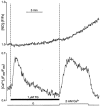
Simultaneous recordings of [Ca2+]i (bottom trace) and [NO]i (top trace) from CPAE cells loaded with fura-2 and DAF-2. Intracellular Ca2+ stores were depleted by exposure to thapsigargin (TG, 1 μm) in the absence of extracellular Ca2+. CCE was elicited by increasing [Ca2+]o to 2 mm.
Figure 4
Figure 4. Inhibition of ATP-induced CCE by NO
A, simultaneous recordings of [Ca2+]i (bottom trace) and [NO]i (top trace) from CPAE cells. Cells were exposed to ATP (5 μm) in the absence of extracellular Ca2+ to deplete intracellular Ca2+ stores. After recovery of the ATP-induced [Ca2+]i transient, 2 mm Ca2+ was added to induce CCE. SNP, added 120 s before Ca2+ application, significantly inhibited ATP-induced CCE. B, the rate of Mn2+ entry ([Mn2+]o= 100 μm; [Ca2+]o= 0) was monitored as the rate of quenching of fura-2 fluorescence excited at the Ca2+-insensitive (isosbestic) wavelength of 360 nm (F360; normalized). The fast Mn2+ influx triggered by ATP stimulation was significantly suppressed by SNP (100 μm). The Mn2+ quench F360 trace labelled ‘-SNP’ was obtained from a cell stimulated with ATP but not subsequently exposed to SNP.
Figure 6
Figure 6. Effect of NO on CCE parameters after store depletion with thapsigargin and ATP
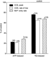
Summary of the effects of SNP on CCE parameters after store depletion with ATP stimulation (left) and thapsigargin (TG) exposure (right). The columns represent the amplitude of the CCE transient (filled column; measured 1 min after addition of Ca2+), the rate of rise of [Ca2+]i during the CCE transient (open column) and the rate Mn2+ entry measured as the rate of quenching of fura-2 fluorescence (hatched column). The data are presented as average percentages ±s.e.m. of control, i.e. the same parameters measured in the absence of SNP. Numbers in parentheses indicate the number of individual cells. The rate of rise of [Ca2+]i and the rate of Mn2+ entry were measured as the slope of the linear portion of the signals presented in Figs 4 and 5.
Figure 5
Figure 5. Effect of NO on CCE triggered by thapsigargin-induced store depletion
A, CCE was triggered by exposure of CPAE cells to 2 mm Ca2+ (filled bar). Intracellular Ca2+ stores were depleted previously (not shown) by exposure to thapsigargin (TG, 1 μm) for 15 min in the absence of extracellular Ca2+. Activation of CCE in the presence of SNP inhibited CCE, resulting in a slower rate of rise of [Ca2+]i, a lower amplitude and slower decline of the CCE transient. B, addition of Mn2+ (100 μm) to thapsigargin-pretreated cells stimulated a fast Mn2+ influx, which was partially suppressed by SNP ([Ca2+]o= 0). The control Mn2+ quench F360 trace labelled ‘-SNP’ was obtained from a thapsigargin-treated cell not subsequently exposed to SNP.
Figure 7
Figure 7. Inhibition of CCE by SNP is mediated by NO and cGMP
CCE transients elicited with 2 mm extracellular Ca2+ after store depletion with thapsigargin (1 μm). Changes of [Ca2+]i evoked by thapsigargin itself are not shown. A, pretreament of CPAE cells with the NO scavenger haemoglobin (Hb) abolished the inhibitory effect of SNP on CCE. Hb (10 μm) was added 100 s before SNP application. B, preincubation with the membrane-permeant cGMP analogue 8-bromo-cGMP (300 μm) before addition of 2 mm Ca2+ inhibited CCE. C, CCE transient in the absence and presence of SNP (100 μm) and 8-bromo-cGMP (300 μm). D, effect of SNP on CCE in the presence of the NO-sensitive guanylate cyclase inhibitor ODQ (10 μm). Cells were preincubated for 40 min.
Figure 8
Figure 8. Inhibition of the PMCA by NO
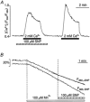
A, CCE transients elicited by brief (< 50 s) exposure to 2 mm extracellular Ca2+ in the absence and presence of SNP (100 μm). Extracellular Ca2+ was removed again during the rising phase of the CCE transient. Intracellular Ca2+ stores were previously depleted by thapsigargin treatment. B, normalized decaying phases of the CCE transients (taken from Figs 5A and 7B) after removal of extracellular Ca2+ under control conditions and in the presence of SNP and 8-bromo-cGMP (cGMP), respectively. The [Ca2+]i signals were normalized to the level of [Ca2+]i encountered at the time of removal of extracellular Ca2+. C, inhibition of the PMCA by lanthanum ([La3+]= 1 mm). La3+ was applied when Ca2+ was removed from the extracellular solution. D, inhibition of the PMCA by La3+ in the presence of SNP. E, average normalized time constants (τ) of the decay phase of the CCE transients under control conditions (100 %) and in the presence of SNP, 8-bromo-cGMP (cGMP), La3+ and La3++ SNP. τ was determined by a monoexponential fit to the declining phase of the CCE transient. The numbers in parentheses indicate the number of cells for each group.
Figure 9
Figure 9. Effect of SNP on the amount of releasable Ca2+ from ionomycin-sensitive Ca2+ stores
Ionomycin (Iono, 1 μm) was applied in the absence of extracellular Ca2+ at the end of each experiment, to estimate the amount of releasable Ca2+ from intracellular stores. A, control [Ca2+]i transient due to ionomycin-induced Ca2+ release. B, stimulation with ATP (5 μm) prior to ionomycin exposure decreased the releasable amount of Ca2+. C, ionomycin treatment following ATP stimulation in the presence of SNP (100 μm, applied 200 s prior to ATP stimulation). The presence of SNP reduced the amplitude of the ATP-induced [Ca2+]i transient, accelerated its recovery and augmented the amount of store Ca2+ releasable by ionomycin exposure. D, ionomycin-releasable [Ca2+]i in a CPAE cell with disabled ER. ER Ca2+ release was abolished by preceding exposure to thapsigargin (TG, 1 μm for 15 min) and verified by the lack of a response to ATP stimulation. E, ionomycin-induced [Ca2+]i signal in a thapsigargin-treated cell in the presence of SNP. Panels A to E are from different cells.
Figure 10
Figure 10. Effect of SNP on Ca2+ reuptake into the stores
A, fura-2-loaded CPAE cells were stimulated twice with ATP (5 μm) in the absence of extracellular Ca2+. Between ATP stimulations, Ca2+ stores were allowed to refill in the presence of 2 mm extracellular Ca2+ for 20 min. B, ATP stimulation in the presence of SNP. SNP was introduced for 1 min prior to the second ATP stimulation; 20 min was allowed for refilling of the stores between ATP stimulations. C, ATP stimulation after store loading in the presence of SNP. SNP was present during the entire 20 min reloading period between the two ATP stimulations.
Figure 11
Figure 11. NO-dependent enhancement of Ca2+ loading of the ER revealed by direct measurements of store content with the low-affinity indicator mag-fura-2
CPAE cells were loaded with mag-fura-2 AM and BAPTA AM. Changes in store [Ca2+] are expressed as changes of the fluorescence ratio F340/F380, reflecting Ca2+ movements in and out of intracellular Ca2+ stores. A, stimulation of cells with ATP (5 μm) in Ca2+-containing medium evoked release of Ca2+ from the stores, resulting in a rapid decline of the mag-fura-2 ratio. Removal of ATP initiated reuptake of Ca2+ into stores. After a 20 min refilling period, accumulated Ca2+ was released by a second stimulation with ATP. B, SNP (100 μm) application during the 20 min refilling period after the first stimulation with ATP resulted in enhanced store loading. A subsequent second challenge with ATP resulted in enhanced Ca2+ release and acceleration of Ca2+ reuptake.

Similar articles

Cited by

References

    1. Assreuy J, Cunha FQ, Liew FY, Moncada S. Feedback inhibition of nitric oxide synthase activity by nitric oxide. British Journal of Pharmacology. 1993;108:833–837. - PMC - PubMed
    1. Berkels R, Dachs C, Roesen R, Klaus W. Simultaneous measurement of intracellular Ca2+ and nitric oxide: A new method. Cell Calcium. 2000a;25:281–286. - PubMed
    1. Berkels R, Suerhoff S, Roesen R, Klaus W. Nitric oxide causes a cGMP-independent intracellular calcium rise in porcine endothelial cells - a paradox? Microvascular Research. 2000b;59:38–44. - PubMed
    1. Bischof G, Serwold TS, Machen TE. Does nitric oxide regulate capacitative Ca2+ influx in HEK 293 cells? Cell Calcium. 1997;21:135–142. - PubMed
    1. Blatter LA, Wier WG. Nitric oxide decreases [Ca2+]i in vascular smooth muscle by inhibition of the calcium current. Cell Calcium. 1994;15:122–131. - PubMed

Publication types

MeSH terms

LinkOut - more resources