. 2002 Feb;109(4):509-16.
doi: 10.1172/JCI14270.

Role of sarcolemmal K(ATP) channels in cardioprotection against ischemia/reperfusion injury in mice

Affiliations

Role of sarcolemmal K(ATP) channels in cardioprotection against ischemia/reperfusion injury in mice

Masashi Suzuki et al. J Clin Invest. 2002 Feb.

Abstract

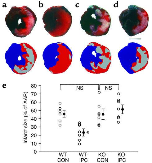
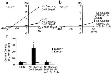
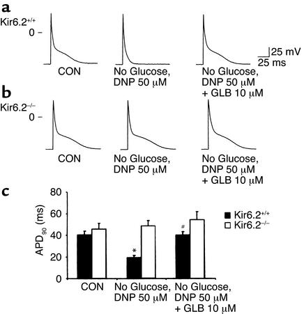
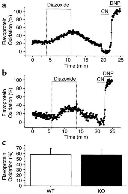
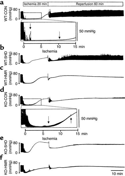
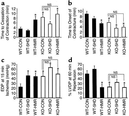
Recently it has been postulated that mitochondrial ATP-sensitive K(+) (mitoK(ATP)) channels rather than sarcolemmal K(ATP) (sarcK(ATP)) channels are important as end effectors and/or triggers of ischemic preconditioning (IPC). To define the pathophysiological significance of sarcK(ATP) channels, we conducted functional experiments using Kir6.2-deficient (KO) mice. Metabolic inhibition with glucose-free, dinitrophenol-containing solution activated sarcK(ATP) current and shortened the action potential duration in ventricular cells isolated from wild-type (WT) but not KO mice. MitoK(ATP) channel function was preserved in KO ventricular cells. In anesthetized mice, IPC reduced the infarct size in WT but not KO mice. Following global ischemia/reperfusion, the increase of left ventricular end-diastolic pressure during ischemia was more marked, and the recovery of contractile function was worse, in KO hearts than in WT hearts. Treatment with HMR1098, a sarcK(ATP) channel blocker, but not 5-hydroxydecanoate, a mitoK(ATP) channel blocker, produced a deterioration of contractile function in WT hearts comparable to that of KO hearts. These findings suggest that sarcKATP channels figures prominently in modulating ischemia/reperfusion injury in the mouse. The rapid heart rate of the mouse (>600 beats per minute) may magnify the relative importance of sarcK(ATP) channels during ischemia, prompting caution in the extrapolation of the conclusions to larger mammals.


Figures

Figure 1
Figure 1
In vivo myocardial infarction studies. (ad) Representative photographs of myocardial slices of WT-CON mice (a), WT-IPC mice (b), KO-CON mice (c), and KO-IPC mice (d) are shown. A schema of each photograph is indicated below; infarct area is expressed as gray, viable myocardium in AAR as red, and nonischemic area as blue. Scale bar = 2 mm. (e) Myocardial infarct size expressed as percentage of AAR for WT-CON (n = 6), WT-IPC (n = 6), KO-CON (n = 6), and KO-IPC (n = 6). There were no significant differences in the infarct size between WT-CON and KO-CON or between KO-CON and KO-IPC. Values are expressed as mean ± SE. NS, not significant. *P < 0.01 versus WT-CON.
Figure 2
Figure 2
Effects of metabolic inhibition with a glucose-free, DNP-containing (50 μM) solution and coapplication of glibenclamide (GLB; 10 μM) on the whole-cell membrane currents recorded from ventricular cells of WT (a) and KO mice (b). (c) Current densities at 0 mV in WT (n = 10) and KO ventricular cells (n = 10) are summarized. Values are expressed as mean ± SE. *P < 0.01 versus control (CON); #P < 0.01 versus DNP, no glucose.
Figure 3
Figure 3
Effects of metabolic inhibition with a glucose-free, DNP-containing (50 μM) solution and coapplication of glibenclamide (GLB; 10 μM) on the action potentials recorded from ventricular cells of WT (a) and KO mice (b). (c) Summarized changes of APD90 in WT (n = 8) and KO cells (n = 8). Values are expressed as mean ± SE. *P < 0.01 versus control (CON); #P < 0.01 versus DNP, no glucose.
Figure 4
Figure 4
Mitochondrial KATP channel activities assessed by changes in flavoprotein fluorescence. The effects of 100 μM diazoxide on flavoprotein fluorescence of representative cells isolated from (a) WT and (b) KO mice. Bars indicate periods of diazoxide application. (c) Summarized data for percentage of flavoprotein oxidation. Despite the absence of surface IK,ATP in KO cells, they displayed response to diazoxide similar to that of WT.
Figure 5
Figure 5
Changes in LVP during ischemia and reperfusion. Representative LVP changes in untreated WT (WT-CON, a), 5-HD–treated WT (WT-5HD, b), HMR1098-treated WT (WT-HMR, c), untreated KO (KO-CON, d), 5-HD–treated KO (KO-5HD, e), and HMR1098-treated KO hearts (KO-HMR, f) are shown. The traces from WT-CON and KO-CON hearts are partly expanded in the insets. Arrows with full line and those with dashed line show the time points of cessation of contraction and onsets of contracture, respectively. Note that KO hearts continued to contract for a longer time than the WT hearts did during ischemia.
Figure 6
Figure 6
Summarized data of mechanical function of isolated hearts during ischemia/reperfusion. Time to cessation of contraction (a), time to onset of contracture (b), EDP at 15 minutes of ischemia (c), and LVDP at 60 minutes of reperfusion (d) in untreated WT (WT-CON, n = 7), 5-HD–treated WT (WT-5HD, n = 7), HMR1098-treated WT (WT-HMR, n = 7), control KO (KO-CON, n = 7), 5-HD–treated KO (KO-5HD, n = 7), and HMR1098-treated KO hearts (KO-HMR, n = 7) are shown. Values are expressed as mean ± SE. *P < 0.05 versus WT-CON.

References

    1. Murry CE, Jennings RB, Reimer KA. Preconditioning with ischemia: a delay of lethal cell injury in ischemic myocardium. Circulation. 1986;74:1124–1136. - PubMed
    1. Liu GC, Vasquez JA, Gallagher KP, Lucchesi BR. Myocardial protection with preconditioning. Circulation. 1990;82:609–619. - PubMed
    1. Scott RJ, Rohmann S, Braun ER, Schaper W. Ischemic preconditioning reduces infarct size in swine myocardium. Circ Res. 1990;66:1133–1142. - PubMed
    1. Bolli R. The early and late phases of preconditioning against myocardial stunning and the essential role of oxyradicals in the late phase: an overview. Basic Res Cardiol. 1996;91:51–63. - PubMed
    1. Reimer KA, Murry CE, Yamasawa I, Hill ML, Jennings RB. Four brief periods of myocardial ischemia cause no cumulative ATP loss or necrosis. Am J Physiol. 1986;251:H1306–H1315. - PubMed

Publication types

MeSH terms