. 2002 Feb 18;195(4):507-16.
doi: 10.1084/jem.20011666.

Virus-induced interferon alpha production by a dendritic cell subset in the absence of feedback signaling in vivo

Affiliations

Virus-induced interferon alpha production by a dendritic cell subset in the absence of feedback signaling in vivo

Winfried Barchet et al. J Exp Med. .

Abstract

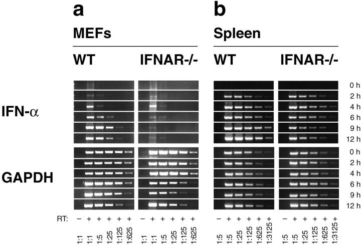
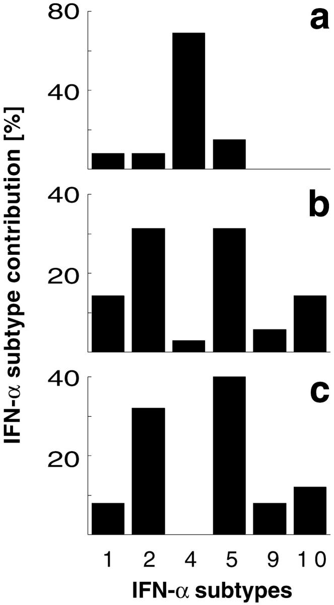
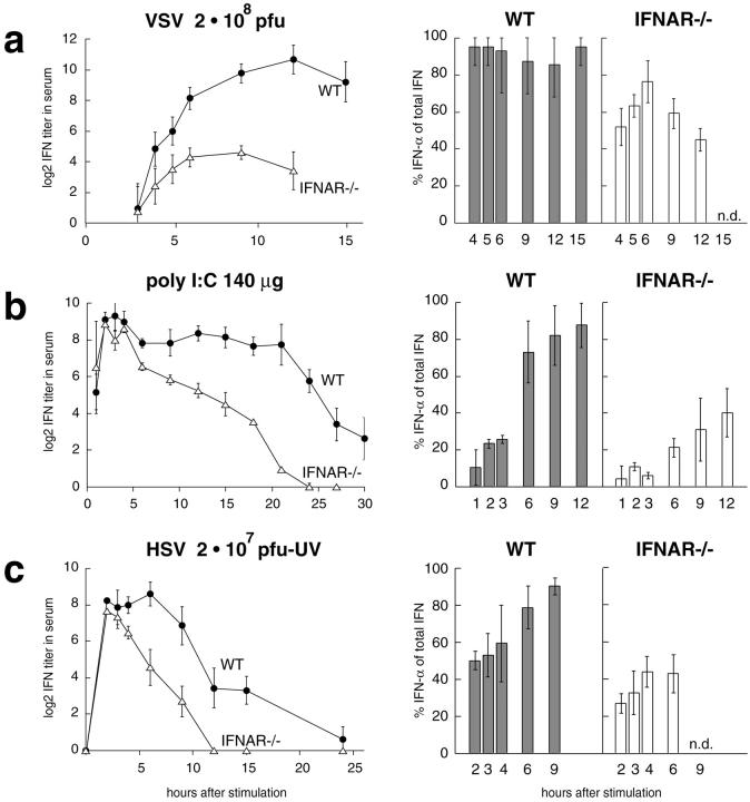
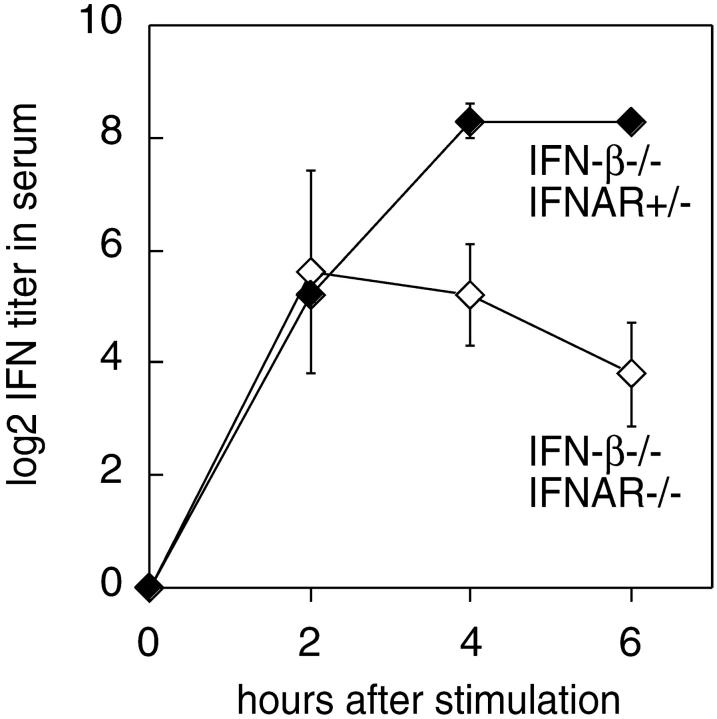
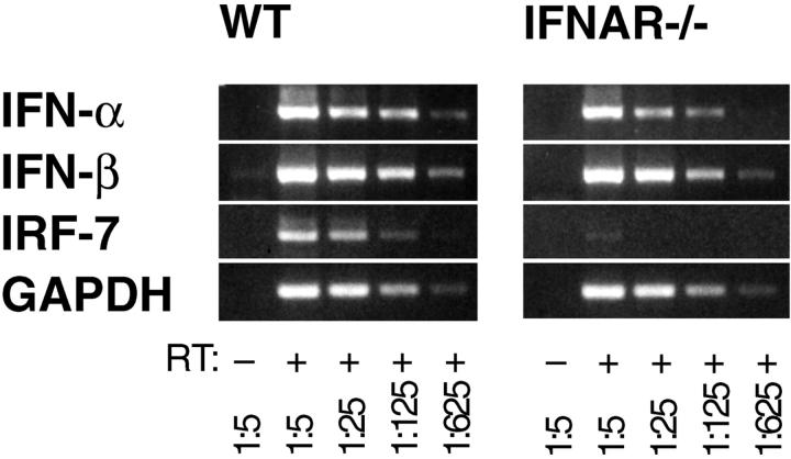
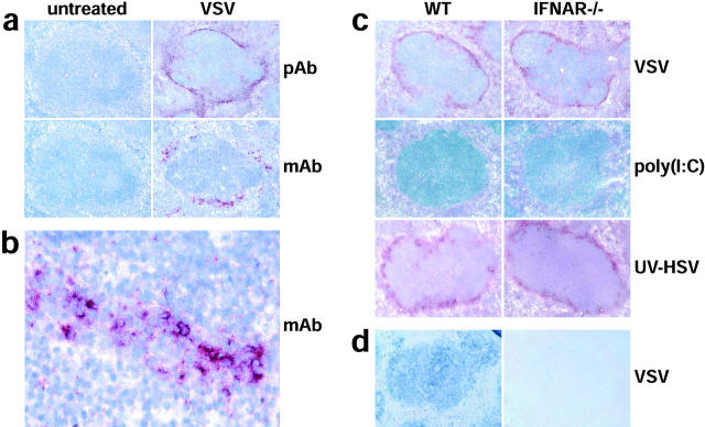
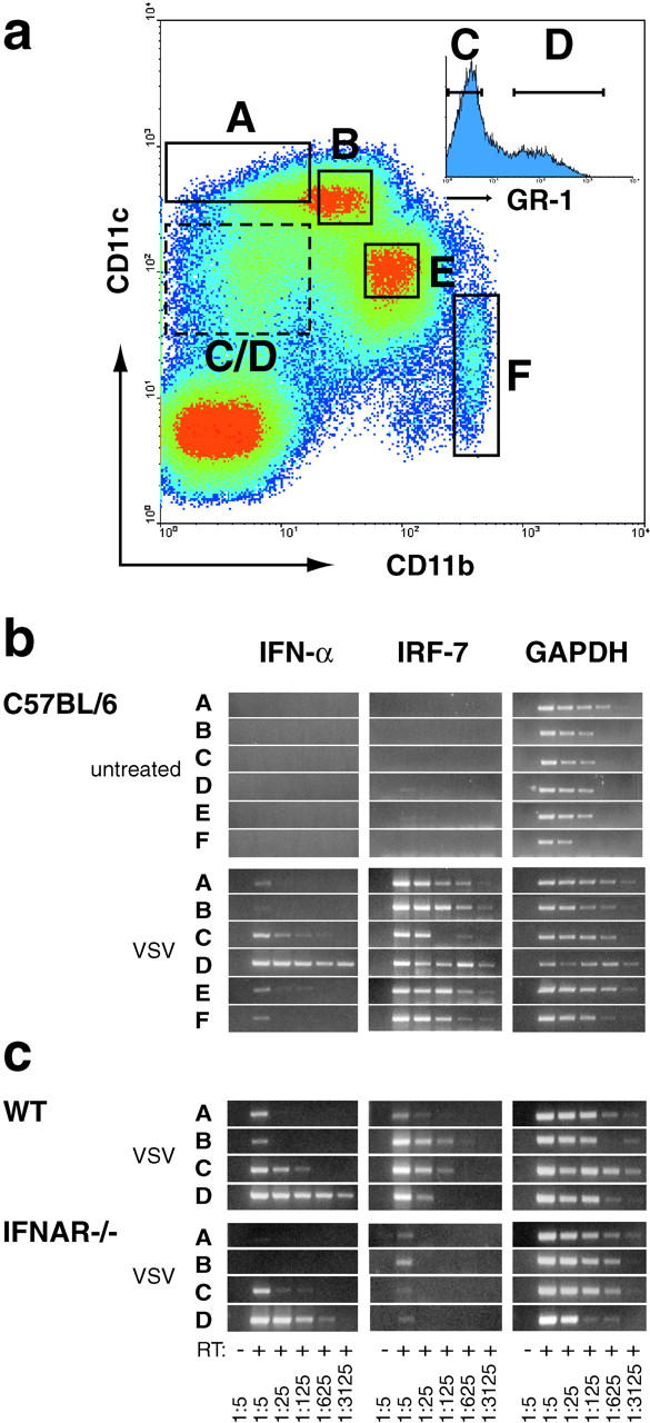
An effective type I interferon (IFN-alpha/beta) response is critical for the control of many viral infections. Here we show that in vesicular stomatitis virus (VSV)-infected mouse embryonic fibroblasts (MEFs) the production of IFN-alpha is dependent on type I IFN receptor (IFNAR) triggering, whereas in infected mice early IFN-alpha production is IFNAR independent. In VSV-infected mice type I IFN is produced by few cells located in the marginal zone of the spleen. Unlike other dendritic cell (DC) subsets, FACS((R))-sorted CD11c(int)CD11b(-)GR-1(+) DCs show high IFN-alpha expression, irrespective of whether they were isolated from VSV-infected IFNAR-competent or -deficient mice. Thus, VSV preferentially activates a specialized DC subset presumably located in the marginal zone to produce high-level IFN-alpha largely independent of IFNAR feedback signaling.


Figures

Figure 1.
Figure 1.
VSV infection stimulates IFNAR-independent production of IFN-α in mice, but not in MEFs. (a) MEFs derived from WT or IFNAR-deficient mice (IFNAR−/−) were infected with VSV at a MOI of 10 and (b) WT and IFNAR−/− mice were intravenously injected with 2 × 10⁸ PFU VSV. Total RNA of 106 cultured MEFs or of spleen tissue was extracted, and the mRNA content of different samples was normalized by a GAPDH-specific RT-PCR. The expression of IFN-α mRNA was monitored using consensus primers amplifying all IFN-α subtypes. The analysis was performed with serially fivefold diluted cDNA samples starting with undiluted material; spleen derived samples were fivefold prediluted.
Figure 2.
Figure 2.
Differential IFN-α expression profiles of VSV infected MEFs and mice. PCR products of the IFN-α expression analysis 6 h after VSV infection of (a) WT MEFs, (b) spleen of WT mice, or (c) spleen of IFNAR−/− mice were subcloned and DNA sequence of 13, 35, and 25 single clones was analyzed, respectively. IFN-α sequences were classified according to EMBL database entries. The sequence termed IFN-α10 corresponds to the IFN-α/β sequence available on the EMBL database (GenBank/EMBL/DDBJ accession no. L38698).
Figure 3.
Figure 3.
Type I IFN activity in the serum of mice treated with VSV, poly(I:C), or UV-HSV. WT mice (black circles) and IFNAR−/− (white triangles) mice were intravenously stimulated with (a) 2 × 10⁸ PFU VSV, (b) 100 μg poly(I:C), or (c) 2 × 10⁷ PFU UV-HSV. Mice were bled at the indicated time points and serum IFN titers were determined in a CPE protection assay. A log2 of 5 corresponds to 1,000 IU of IFN-α. The contribution of IFN-α or IFN-β to the total IFN activity was determined by specific inhibition with mAbs.
Figure 4.
Figure 4.
IFN-α is produced in mice deficient of IFN-β and IFNAR. IFN-β–deficient mice (black diamonds) and double-deficient mice lacking IFN-β and IFNAR (white diamonds) were intravenously injected with 2 × 10⁷ PFU UV-HSV. Sera were taken at indicated time points and analyzed in a CPE protection assay. Mean IFN activity of three mice per time point is indicated. Results are shown of one out of two independent experiments. Note that the indicated IFN serum activities exclusively derived form IFN-α because the analyzed mice were IFN-β deficient.
Figure 5.
Figure 5.
VSV infected IFNAR−/− mice show an early induction of IFN-α before the onset of IRF-7 expression. Total RNA from spleen of IFNAR−/− or WT mice was isolated 3 h after intravenous infection with 2 × 10⁸ PFU VSV. RT-PCR was performed as described in Fig. 1.
Figure 6.
Figure 6.
VSV and UV-HSV injection, but not poly(I:C) stimulation, leads to high-level production of type I IFN by cells located in the marginal zone of the spleen. (a) WT mice were VSV infected and after 9 h spleens were analyzed immunohistochemically with a polyclonal serum against type I IFN (pAb, top two panels) or with an IFN-α–specific mAb (mAb, bottom two panels). (b) A higher magnification of the marginal zone area of the bottom right panel in (a) stained with mAb is shown. (c) WT and IFNAR−/− mice were VSV, poly (I:C), or UV-HSV injected. After 9 h and 6 h after UV-HSV stimulation, spleens were analyzed immunohistochemically with a polyclonal serum against type I IFN. Sections in (a–c) were counterstained with hemalum (blue) to visualize lymph follicles. (d) In situ hybridization of IRF-7 mRNA (blue) was performed on paraffin sections of spleen from WT and IFNAR−/− mice prepared 9 h after VSV infection.
Figure 7.
Figure 7.
In VSV-infected mice CD11cintCD11bGR-1+ DCs express high-level IFN-α. (a) Mice were intravenously infected with 2 × 10⁸ PFU VSV and 9 h later spleens were removed to prepare single cell suspensions. MACS®-enriched CD11c+ cells were stained with anti-CD11c-Biot./Str.-APC, anti-CD11b-FITC, and anti-GR-1-PE, and DC subsets were FACS®-sorted using the indicated gates A–F. Cells from gate C/D were further subdivided by separating GR-1 (fraction C) and GR-1+ cells (fraction D). Sorted fractions were derived from spleen of (b) VSV infected or untreated C57BL/6 mice, and of (c) VSV-infected Sv129 (WT) and IFNAR−/− mice. Total RNA of ∼4–10 × 104 sorted cells was prepared and analyzed by RT-PCR as described in Fig. 1.

Comment in

References

    1. Isaacs, A., and J. Lindenmann. 1957. Virus interference. I. The interferon. Proc. R. Soc. Lond. 147:258–267. - PubMed
    1. Gresser, I., M.G. Tovey, C. Maury, and M.T. Bandu. 1976. Role of interferon in the pathogenesis of virus diseases as demonstrated by the use of anti-interferon serum. II. Studies with herpes simplex, Moloney sarcoma, vesicular stomatitis, Newcastle disease, and influenza viruses. J. Exp. Med. 144:1316–1323. - PMC - PubMed
    1. Muller, U., U. Steinhoff, L.F. Reis, S. Hemmi, J. Pavlovic, R.M. Zinkernagel, and M. Aguet. 1994. Functional role of type I and type II interferons in antiviral defense. Science. 264:1918–1921. - PubMed
    1. Cella, M., M. Salio, Y. Sakakibara, H. Langen, I. Julkunen, and A. Lanzavecchia. 1999. Maturation, activation, and protection of dendritic cells induced by double-stranded RNA. J. Exp. Med. 189:821–829. - PMC - PubMed
    1. Le Bon, A., G. Schiavoni, G. D'Agostino, I. Gresser, F. Belardelli, and D.F. Tough. 2001. Type I interferons potently enhance humoral immunity and can promote isotype switching by stimulating dendritic cells in vivo. Immunity. 14:461–470. - PubMed

Publication types

MeSH terms