. 2002 Feb;13(2):503-14.
doi: 10.1091/mbc.01-07-0365.

Pag1p, a novel protein associated with protein kinase Cbk1p, is required for cell morphogenesis and proliferation in Saccharomyces cerevisiae

Affiliations

Pag1p, a novel protein associated with protein kinase Cbk1p, is required for cell morphogenesis and proliferation in Saccharomyces cerevisiae

Li-Lin Du et al. Mol Biol Cell. 2002 Feb.

Abstract

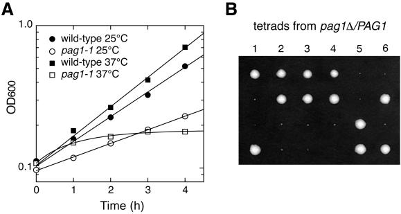
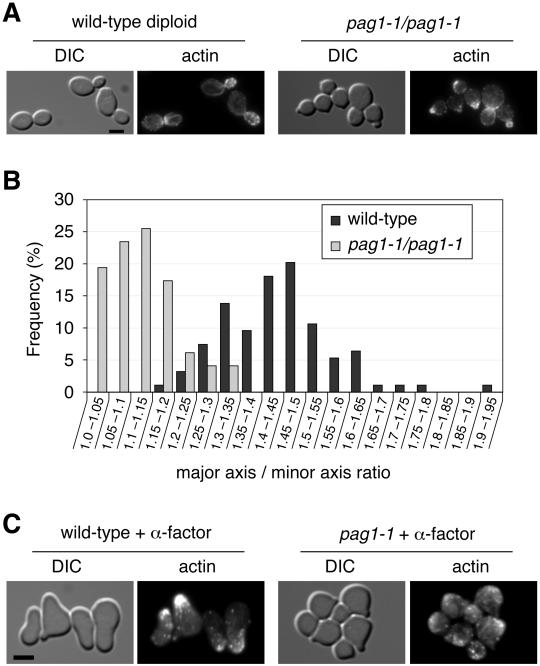
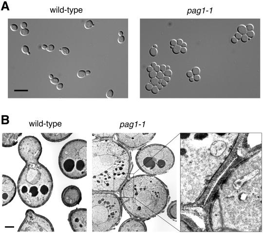
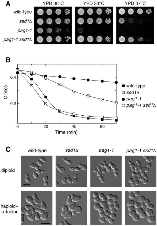
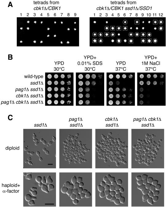
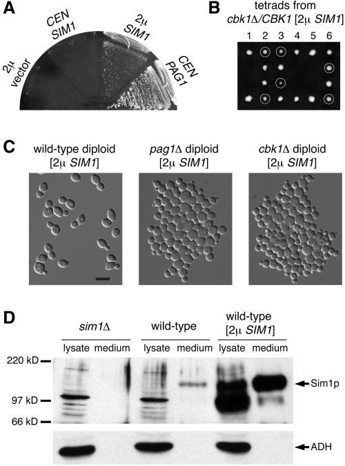
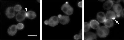
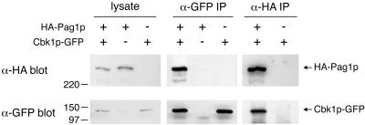
Protein kinases in the Cot-1/Orb6/Ndr/Warts family are important regulators of cell morphogenesis and proliferation. Cbk1p, a member of this family in Saccharomyces cerevisiae, has previously been shown to be required for normal morphogenesis in vegetatively growing cells and in haploid cells responding to mating pheromone. A mutant of PAG1, a novel gene in S. cerevisiae, displayed defects similar to those of cbk1 mutants. pag1 and cbk1 mutants share a common set of suppressors, including the disruption of SSD1, a gene encoding an RNA binding protein, and the overexpression of Sim1p, an extracellular protein. These genetic results suggest that PAG1 and CBK1 act in the same pathway. Furthermore, we found that Pag1p and Cbk1p localize to the same polarized peripheral sites and that they coimmunoprecipitate with each other. Pag1p is a conserved protein. The homologs of Pag1p in other organisms are likely to form complexes with the Cbk1p-related kinases and function with those kinases in the same biological processes.


Figures

Figure 1
Figure 1
PAG1 is essential for growth. (A) Growth properties of the temperature-sensitive mutant pag1-1. Wild-type cells (NY13) or pag1-1 cells (NY2333) growing exponentially at 25°C were shifted to fresh YPD medium at 25 or 37°C. Optical density at 600 nm (OD600) was determined at 1-h intervals. (B) Tetrads derived from the sporulation of pag1Δ/PAG1 (NY2334) were dissected onto a YPD plate and incubated at 25°C for 5 d. Six tetrads are shown. The smaller colonies are pag1Δ cells.
Figure 2
Figure 2
pag1-1 is defective in cell shape and mating projection formation. (A) Wild-type diploid cells (NY1523) and pag1-1 diploid cells (NY2342) growing exponentially in YPD at 30°C were fixed and stained with fluorescently labeled phalloidin. Cells were imaged in differential interference contrast (DIC) mode and fluorescence mode, respectively. Bar, 4 μm. (B) pag1-1 diploid cells are significantly rounder than wild-type cells. From the DIC images, we measured the lengths of the major axis and minor axis of budded cells of the two strains shown in A. Distributions of the major axis/minor axis ratio are plotted as a histogram. (C) Wild-type MATa cells (NY13) and pag1-1 MATa cells (NY2333) grown in YPD at 30°C were treated with α-factor (5 μg/ml) at 30°C for 3 h then fixed and stained with fluorescently labeled phalloidin. Bar, 4 μm.
Figure 3
Figure 3
pag1-1 cells have a separation defect. (A) pag1-1 cells form aggregates in liquid culture. Wild-type cells (NY13) and pag1-1 cells (NY2333) growing exponentially in YPD at 25°C were examined by light microscopy. Bar, 8 μm. (B) Electron microscopy shows that aggregated pag1-1 cells are connected by septa. Bar, 1 μm. A septum between two pag1-1 cells is shown at a higher magnification at right.
Figure 4
Figure 4
Deletion of SSD1 strongly suppresses the growth defect of pag1-1 but does not significantly rescue the defects in cell morphogenesis. (A) ssd1Δ suppresses the growth defect of pag1-1 and its sensitivity to SDS and 1 M NaCl. Tenfold serial dilutions of wild-type (NY2337), ssd1Δ (NY2344), pag1-1 (NY2343), and pag1-1 ssd1Δ (NY2345) cells were spotted on plates and incubated at the indicated temperatures. Photos were taken 40 h later. (B) pag1-1 is resistant to β-1,3-glucanase treatment and ssd1Δ partially suppresses this phenotype. The same yeast strains as used in A were grown in YPD at 30°C and then treated with β-1,3-glucanase (10 U/ml Quantazyme ylg). Cell lysis was monitored by the decrease of OD600. (C) ssd1Δ does not significantly suppress the defects of pag1-1 in cell separation and polarized growth. Wild-type (NY2349), ssd1Δ (NY2351), pag1-1 (NY2362), and pag1-1 ssd1Δ (NY2363) homozygous diploid cells growing exponentially in YPD at 30°C were fixed and photographed. The same MATa strains as used in A were grown in YPD at 30°C, treated with α-factor (5 μg/ml) for 3 h at 30°C, and then fixed and photographed. Bar, 8 μm.
Figure 5
Figure 5
cbk1Δ ssd1Δ cells display the same phenotype as pag1Δ ssd1Δ cells. (A) The lethality of cbk1Δ in the genetic background of the genome deletion project can be suppressed by ssd1Δ. Tetrads derived from cbk1Δ/CBK1 (NY2350) and cbk1Δ/CBK1 ssd1Δ/SSD1 (NY2361) diploid cells were dissected onto YPD plates and incubated at 25°C. Photos were taken 4 d later. The genotypes of the haploid progeny were determined by PCR analysis. ssd1Δ colonies are circled. cbk1Δ ssd1Δ colonies are enclosed in squares. Unmarked colonies are wild-type. (B) pag1Δ ssd1Δ, cbk1Δ ssd1Δ, and pag1Δ cbk1Δ ssd1Δ cells are equally hypersensitive to SDS and NaCl. Tenfold serial dilutions of haploid cells were spotted on plates and incubated at the indicated temperatures. Photos were taken 40 h later for the YPD plates and the YPD plate containing SDS and 90 h later for the YPD plate containing NaCl. (C) pag1Δ ssd1Δ, cbk1Δ ssd1Δ, and pag1Δ cbk1Δ ssd1Δ cells display similar defects in cell separation and polarized growth. The homozygous diploid cells growing exponentially in YPD at 30°C were fixed and photographed. The MATa strains grown in YPD at 30°C were treated with α-factor (5 μg/ml) for 3 h at 30°C and then fixed and photographed. Bar, 8 μm.
Figure 6
Figure 6
Overexpression of an extracellular protein, Sim1p, suppresses the growth defects of both pag1Δ and cbk1Δ. (A) SIM1 is a dosage-dependent suppressor of pag1Δ. pag1Δ [2 μ URA3 PAG1] (NY2336) with the HIS3-containing plasmids shown in the figure streaked onto a 5-FOA plate and incubated at 25°C. (B) A multicopy [2 μ URA3 SIM1] plasmid suppresses the lethality of cbk1Δ. Tetrads were dissected onto a YPD plate and incubated at 25°C. Photo was taken 4 d later. G-418 resistant colonies (cbk1Δ) are circled. They are Ura+ and cannot grow on a 5-FOA plate. (C) pag1Δ and cbk1Δ cells with a multicopy SIM1 plasmid display defects in cell separation and polarized growth. Wild-type (NY2355), pag1Δ (NY2356), and cbk1Δ (NY2357) homozygous diploid cells containing a [2 μ URA3 SIM1] plasmid growing exponentially in SC-URA at 30°C were fixed and photographed. (D) Sim1p is a secreted protein. Cells were grown to log phase in synthetic media. Lysates were made by vortexing with glass beads. Proteins secreted into the medium were precipitated with trichloroacetic acid. Samples corresponding to materials from cultures containing 0.4 OD600 unit cells were separated by SDS-PAGE. Western blot was probed with antibodies against Sim1p and a cytosolic protein, alcohol dehydrogenase (ADH).
Figure 7
Figure 7
Pag1p exhibits a polarized localization to the cell periphery. Localization of GFP-tagged Pag1p was detected in living cells (NY2335) grown in YPD. GFP-Pag1p expressed from the PAG1 promoter was the sole copy of the full-length Pag1p in this strain. Bar, 4 μm. A mother-daughter neck and a presumptive budding site are indicated by an arrowhead and an arrow, respectively.
Figure 8
Figure 8
HA-tagged Pag1p coimmunoprecipitates with GFP-tagged Cbk1p. Lysates were prepared from yeast cells expressing HA-Pag1p, Cbk1p-GFP or both. Lysates and immunoprecipitates with anti-HA and anti-GFP antibodies were separated by SDS-PAGE and probed with anti-HA and anti-GFP antibodies.

References

    1. Adams A, Gottschling DE, Kaiser CA, Stearns T. Methods in Yeast Genetics. Cold Spring Harbor, NY: Cold Spring Harbor Laboratory; 1997.
    1. Bender A, Pringle JR. Use of a screen for synthetic lethal and multicopy suppressee mutants to identify two new genes involved in morphogenesis in Saccharomyces cerevisiae. Mol Cell Biol. 1991;11:1295–1305. - PMC - PubMed
    1. Bidlingmaier S, Weiss EL, Seidel C, Drubin DG, Snyder M. The Cbk1p pathway is important for polarized cell growth and cell separation in Saccharomyces cerevisiae. Mol Cell Biol. 2001;21:2449–2462. - PMC - PubMed
    1. Cong J, Geng W, He B, Liu J, Charlton J, Adler PN. The furry gene of Drosophila is important for maintaining the integrity of cellular extensions during morphogenesis. Development. 2001;128:2793–2802. - PubMed
    1. Cormack BP, Bertram G, Egerton M, Gow NA, Falkow S, Brown AJ. Yeast-enhanced green fluorescent protein (yEGFP) Microbiology. 1997;143:303–311. - PubMed

Publication types

MeSH terms

Substances