. 2002 Feb;13(2):530-41.
doi: 10.1091/mbc.01-09-0440.

Secretion and assembly of zona pellucida glycoproteins by growing mouse oocytes microinjected with epitope-tagged cDNAs for mZP2 and mZP3

Affiliations

Secretion and assembly of zona pellucida glycoproteins by growing mouse oocytes microinjected with epitope-tagged cDNAs for mZP2 and mZP3

Huayu Qi et al. Mol Biol Cell. 2002 Feb.

Abstract

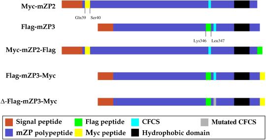
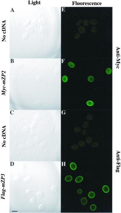
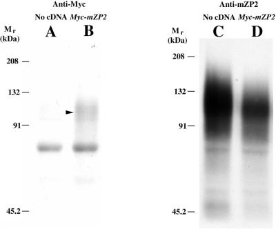
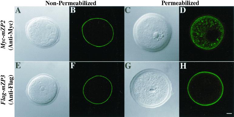
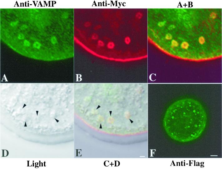
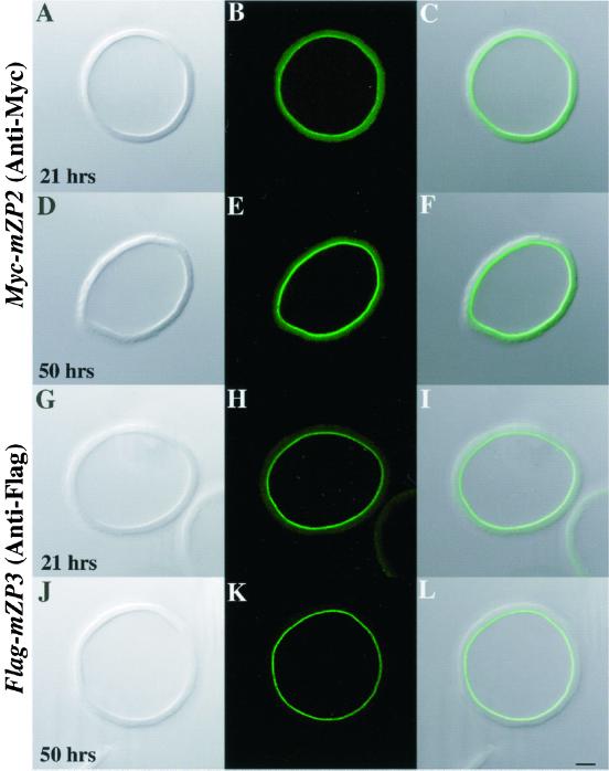
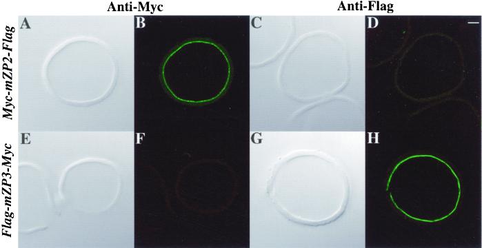
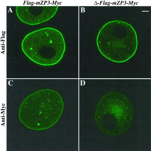
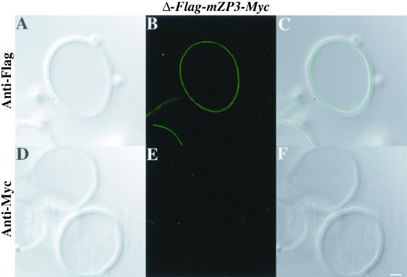
The zona pellucida (ZP) is a highly organized extracellular coat that surrounds all mammalian eggs. The mouse egg ZP is composed of three glycoproteins, called mZP1-3, that are synthesized, secreted, and assembled into a ZP exclusively by growing oocytes. Here, we microinjected epitope-tagged (Myc and Flag) cDNAs for mZP2 and mZP3 into the germinal vesicle (nucleus) of growing oocytes isolated from juvenile mice. Specific antibodies and laser scanning confocal microscopy were used to follow nascent, recombinant ZP glycoproteins in both permeabilized and nonpermeabilized oocytes. When such cDNAs were injected, epitope-tagged mZP2 (Myc-mZP2) and mZP3 (Flag-mZP3) were synthesized, packaged into large intracellular vesicles, and secreted by the vast majority of oocytes. Secreted glycoproteins were incorporated into only the innermost layer of the thickening ZP, and the amount of nascent glycoprotein in this region increased with increasing time of oocyte culture. Consistent with prior observations, the putative transmembrane domain at the C terminus of mZP2 and mZP3 was missing from nascent glycoprotein incorporated into the ZP. When the consensus furin cleavage site near the C terminus of mZP3 was mutated, such that it should not be cleaved by furin, secretion and assembly of mZP3 was reduced. On the other hand, mZP3 incorporated into the ZP lacked the transmembrane domain downstream of the mutated furin cleavage site, suggesting that some other protease(s) excised the domain. These results strongly suggest that nascent mZP2 and mZP3 are incorporated into only the innermost layer of the ZP and that excision of the C-terminal region of the glycoproteins is required for assembly into the oocyte ZP.


Figures

Figure 1
Figure 1
Schematic representation of polypeptides encoded by cDNA constructs microinjected into growing mouse oocytes. Short peptide epitopes, Myc (yellow) and Flag (green), were inserted into mZP2 and mZP3 polypeptides. In singly tagged ZP glycoproteins, Myc (-Glu-Gln-Lys-Leu-Ile-Ser-Glu-Glu-Asp-Leu-) was inserted into mZP2 (Myc-mZP2), after the N-terminal signal sequence, between amino acids Gln-39 and Ser-40. Flag (-Asp-Tyr-Lys-Asp-Asp-Asp-Asp-Lys-) was inserted into mZP3 (Flag-mZP3) near its C terminus, before the CFCS, between amino acids Lys-346 and Leu-347. In doubly tagged ZP glycoproteins, epitope tags were inserted at the C terminus of Myc-mZP2 and Flag-mZP3, producing Myc-mZP2-Flag and Flag-mZP3-Myc, respectively. Also shown is Δ-Flag-mZP3-Myc, a construct in which the CFCS of mZP3 is mutated from -Arg-Asn-Arg-Arg- to -Arg-Asn-Gly-Glu- (gray). Several features of mZP2 and mZP3, such as the positions of the signal sequence (red), CFCS (royal blue), and transmembrane (hydrophobic) domain (black), are indicated.
Figure 2
Figure 2
Immunofluorescence staining of Myc-mZP2 and Flag-mZP3 in growing mouse oocytes. Growing oocytes were isolated from juvenile mice, as described in MATERIALS AND METHODS. After microinjection of Myc-mZP2 and Flag-mZP3 cDNA (∼1 mg/ml) into the oocyte GV, oocytes were cultured for ∼20 h, as described in MATERIALS AND METHODS. Epitope-specific monoclonal antibodies, 9E10 (anti-Myc) and M2 (anti-Flag), were used to immunolabel oocytes at the end of the culture period. Goat anti-mouse IgG coupled to FITC was used as a secondary antibody. Light images of immunolabeled oocytes are presented in A–D and fluorescent images are presented in E–H. Whereas uninjected oocytes showed no staining (A and E [anti-Myc] and C and G [anti-Flag]), injected oocytes exhibited intense fluorescence (B and F [anti-Myc] and D and H [anti-Flag]). The most intense signal was seen at the surface of the oocytes over the plasma membrane/ZP region. In each case, 100–200 oocytes were examined by LSCM. Bar (in D), 50 μm.
Figure 3
Figure 3
Gel analysis of epitope-tagged ZP glycoproteins synthesized and secreted by growing mouse oocytes. Two hundred growing oocytes were isolated from 13-d-old mice, microinjected with Myc-mZP2, and cultured in M199-M for ∼5 h. Oocytes were then transferred to Met/Cys-depleted M199-M, supplemented with 4 mCi/ml [35S]Met/Cys. The culture medium was collected after ∼15 h and immunoprecipitated with monoclonal antibodies directed against Myc (9E10). Immunoprecipitates were subjected to 7.5% SDS-PAGE, as described in MATERIALS AND METHODS. Media from uninjected and injected oocytes probed with anti-Myc are shown in A and B, respectively. An arrowhead indicates the position of mZP2. Media were immunoprecipitated once again with an anti-mZP2 polyclonal antibody. As shown in C and D, both injected and noninjected oocytes secreted endogenous mZP2 glycoproteins. A band at ∼70 kDa was observed as a contaminant in all lanes. The positions of molecular mass standards are shown.
Figure 4
Figure 4
Localization of Myc-mZP2 and Flag-mZP3 in growing mouse oocytes. Oocytes from 14-d-old mice were microinjected with either Myc-mZP2 or Flag-mZP3. After ∼21 h of culture, the oocytes were stained with anti-Myc (A–D) or anti-Flag (E–H), followed by FITC-conjugated secondary antibody. Light (A, C, E, and G) and fluorescent (B, D, F, and H) images of oocytes are presented. In fixed, permeabilized oocytes, both epitope-tagged ZP proteins were detected, primarily over the plasma membrane/ZP region (D and H). In addition, intracellular vesicles were readily observed in Myc-mZP2-injected oocytes (D). Much less intracellular staining was detected in Flag-mZP3-injected oocytes (H), although in some cases, vesicles were detected. The plasma membrane/ZP staining appears to be on the outside of permeabilized growing oocytes, because fixed, nonpermeabilized samples (A, B, E, and F) exhibited very similar staining patterns. In each case, 100–200 oocytes were examined by LSCM. Bar (H), 10 μm.
Figure 5
Figure 5
Colocalization of Myc-mZP2 and VAMP in growing mouse oocytes. Oocytes from 14-d-old mice were microinjected with Myc-mZP2. After ∼21 h of culture, the oocytes were stained with anti-VAMP (IgM; A) and anti-Myc (IgG; B), followed by FITC-conjugated rabbit anti-mouse IgM and Texas Red-conjugated goat anti-mouse IgG secondary antibody. Light (D) and fluorescent (A–C) images of oocytes are presented. C, An overlap of A and B. Note the presence of VAMP (green) and Myc-mZP2 (red) in the large, doughnut-shaped vesicles present in the cortical region of the oocyte; overlap of VAMP and Myc-mZP2 is yellow (green plus red). E, An overlap of C and D. Arrowheads in D and E, the positions of large vesicles in the cortical region of the oocyte. In F, an oocyte microinjected with mZP2 tagged with Flag at its C terminus (Myc-mZP2-Flag) and probed with anti-Flag is shown. Note that the vesicles exhibit fluorescence along their membranes. In each case, 100–200 oocytes were examined by LSCM. Bars (E and F), 2 μm and 10 μm, respectively.
Figure 6
Figure 6
Assembly of Myc-mZP2 and Flag-mZP3 into ZP of growing mouse oocytes. Either Myc-mZP2 (A–F) or Flag-mZP3 (G–L) cDNA was microinjected into growing oocytes isolated from 13-d-old mice. After the oocytes were cultured for ∼21 (A–C and G–I) and ∼50 h (D–F and J–L), ZP were isolated in the presence of 1% NP-40 and immunolabeled with either anti-Myc or anti-Flag and FITC-conjugated secondary antibody. Light images are shown in A, D, G, and J and fluorescent images are shown in B, E, H, and K. Composites of light and fluorescent images are shown in C, F, I, and L. In all cases, epitope-tagged ZP glycoproteins were found only at the innermost layer of the ZP (A–F, Myc-mZP2; G–L, Flag-mZP3). The intensity of the immunofluorescence signal increased with increasing culture times (compare B and E, Myc-mZP2; compare H and K, Flag-mZP3; see text for details). More than 200 isolated ZP were stained in this manner. Bar (L), 10 μm.
Figure 7
Figure 7
Proteolytic processing of epitope-tagged ZP glycoproteins in growing mouse oocytes. Doubly tagged (Myc and Flag) cDNAs for mZP2 and mZP3 were microinjected into growing oocytes isolated from 14-d-old mice. After culture for ∼50 h, ZP were isolated in the presence of 1% NP-40 and stained with either anti-Myc or anti-Flag, followed by FITC-conjugated secondary antibody. Light images are shown in A, C, E, and G and fluorescent images are shown in B, D, F, and H. ZP from Myc-mZP2-Flag-injected oocytes (A–D) exhibited fluorescence only with anti-Myc (B), not with anti-Flag (D). ZP from Flag-mZP3-Myc-injected oocytes (E–H) exhibited fluorescence only with anti-Flag (H), not with anti-Myc (F). Bar (D), 10 μm.
Figure 8
Figure 8
Secretion of epitope-tagged mZP3, mutated at its CFCS, by growing mouse oocytes. Flag-mZP3-Myc and Δ-Flag-mZP3-Myc (mutated at its CFCS) were microinjected into growing oocytes isolated from 14- to 15-d-old mice. Oocytes were cultured for ∼20 h and then stained with either anti-Flag or anti-Myc, followed by FITC-conjugated secondary antibody. Intense fluorescence was evident over the plasma membrane/ZP region of oocytes injected with either cDNA construct after staining with anti-Flag (A and B) but not with anti-Myc (C and D). It was noted that, in almost all instances, there was a larger accumulation of fluorescently labeled vesicles in oocytes microinjected with Δ-Flag-mZP3-Myc (B and D) than in oocytes microinjected with Flag-mZP3-Myc (A and C). In each case, 100–200 oocytes were examined by LSCM. Bar (B), 10 μm.
Figure 9
Figure 9
Assembly of epitope-tagged mZP3, mutated at its consensus furin cleavage site, into the ZP of growing mouse oocytes. Δ-Flag-mZP3-Myc mutated at its CFCS was microinjected into growing oocytes isolated from 14-d-old mice. Oocytes were cultured for ∼50 h, and ZP were isolated in the presence of 1% NP-40 and stained with either anti-Flag or anti-Myc, followed by FITC-conjugated secondary antibody. Light (A and D) and fluorescent (B and E) images are shown for ZP treated with anti-Flag (A–C) or anti-Myc (D–F). C and F represent overlaps of A and B and D and E, respectively. Note that in B (anti-Flag), but not in E (anti-Myc), the innermost layer of the ZP is fluorescently labeled. Bar (F), 10 μm.

Similar articles

Cited by

References

    1. Beebe SL, Leyton L, Burks D, Ishikawa M, Fuerst T, Dean J, Saling P. Recombinant mouse ZP3 inhibits sperm binding and induces the acrosome reaction. Dev Biol. 1992;151:48–54. - PubMed
    1. Bleil JD, Wassarman PM. Autoradiographic visualization of the mouse egg's sperm receptor bound to sperm. J Cell Biol. 1986;102:1363–1371. - PMC - PubMed
    1. Bork P, Sander C. A large domain common to sperm receptors (ZP2 and ZP3) and TGF-β type III receptor. FEBS Lett. 1992;300:237–240. - PubMed
    1. Chalifour LE, Wirak DO, Hansen U, Wassarman PM, DePamphilis ML. Cis- and trans-acting sequences required for the expression of simian virus 40 genes in mouse oocytes. Genes Dev. 1987;1:1096–1106. - PubMed
    1. Chalifour LE, Wirak DO, Wassarman PM, DePamphilis ML. Expression of simian virus 40 early and late genes in mouse oocytes and embryos. J Virol. 1986;59:619–627. - PMC - PubMed

Publication types

MeSH terms

LinkOut - more resources