

doi: 10.1091/mbc.01-07-0336.
The Drosophila nuclear lamina protein YA binds to DNA and histone H2B with four domains
Affiliations
- PMID: 11854412
- PMCID: PMC65649
- DOI: 10.1091/mbc.01-07-0336
Item in Clipboard
The Drosophila nuclear lamina protein YA binds to DNA and histone H2B with four domains
Mol Biol Cell.
2002 Feb.
Abstract
Dramatic changes occur in nuclear organization and function during the critical developmental transition from meiosis to mitosis. The Drosophila nuclear lamina protein YA binds to chromatin and is uniquely required for this transition. In this study, we dissected YA's binding to chromatin. We found that YA can bind to chromatin directly and specifically. It binds to DNA but not RNA, with a preference for double-stranded DNA (linear or supercoiled) over single-stranded DNA. It also binds to histone H2B. YA's binding to DNA and histone H2B is mediated by four domains distributed along the length of the YA molecule. A model for YA function at the end of Drosophila female meiosis is proposed.
Figures
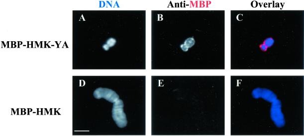
YA binds to mitotic chromosomes directly. Purified MBP-HMK-YA (A–C) or MBP-HMK (D–F) was incubated with mitotic chromosomes. The chromosomes were stained with the DNA dye DAPI (A and D, and blue in C and F) and anti-MBP antisera (B and E, and red in C and F). MBP, maltose binding protein; HMK, phosphorylation sites for heart muscle kinase. The difference in staining between the chromosomes in A–C vs. the one in D–F is due to the difference in the ability of MBP-HMK-YA and MBP-HMK to bind to them; it is not due to their difference in size. Chromosomes of different sizes incubated with the same protein show comparable binding results (e.g., see Figure 8I for two apposed smaller chromosomes incubated with MBP-HMK). Bar: 10 μm.
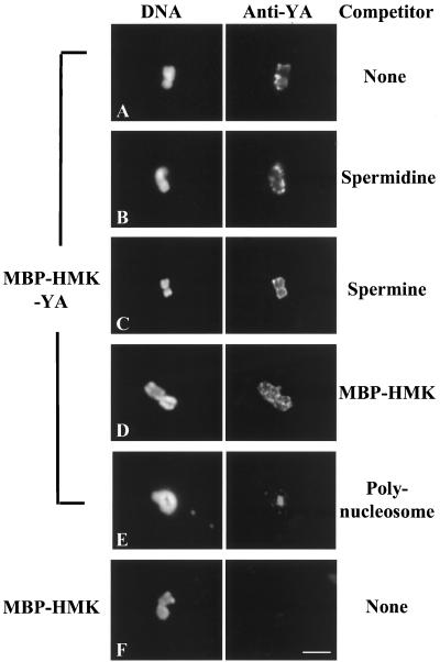
YA's binding to mitotic chromosomes is specific. Binding reactions of MBP-HMK-YA with mitotic chromosomes were carried out in the presence of 1000-fold molar excess of spermidine or spermine, 25-fold molar excess of MBP-HMK or 18-fold molar excess of polynucleosomes. Chromosomes were stained with DAPI (DNA) and anti-YA antibodies (Anti-YA). Bar: 10 μm.
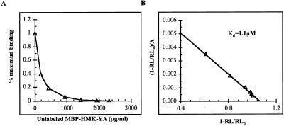
MBP-HMK-YA binds to immobilized chromatin with apparent Kd of 1.1 μM. (A) 32P-labeled MBP-HMK-YA was incubated with immobilized polynucleosomes in the presence of increasing concentration of unlabeled MBP-HMK-YA. Binding at each point was corrected for nonspecific binding by subtraction of values obtained with 2300 μg/ml unlabeled MBP-HMK-YA. (B) Data for specific binding were analyzed as in Hulme and Birdsall (1992). RL is the amount of radioactive protein bound to chromatin at unlabeled protein concentration A. RL0 is the amount of radioactive protein bound to chromatin in absence of unlabeled competitor. The slope given by this plot equals −1/Kd.
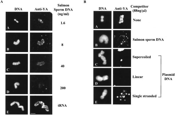
YA's binding to mitotic chromosomes can be competed by salmon sperm DNA, by different forms of plasmid DNA, but not by tRNA. The binding reactions of MBP-HMK-YA with mitotic chromosomes in Figure 4A were performed in the presence of deproteinated salmon sperm DNA at the indicated concentrations (4A, panels A–D) or 200 ng/μl yeast tRNA (4A, panel E). The mitotic chromosome bindings shown in Figure 4B were of 88 ng/μl salmon sperm DNA or plasmid DNA of the indicated forms. Mitotic chromosomes were stained with DAPI (DNA) and anti-YA antibodies (Anti-YA). Bar: 10 μm.
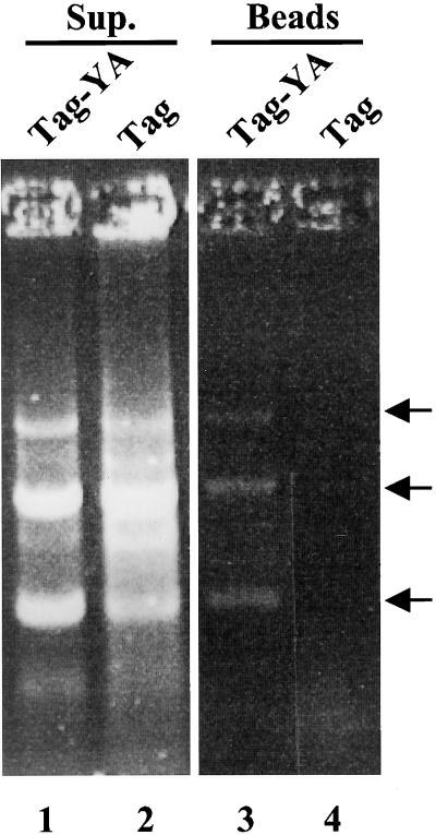
DNA is pulled down with MBP-HMK-YA in a MBP pull-down assay. MBP-HMK-YA (4 μg; Tag-YA) or MBP-HMK (32 μg; Tag) was incubated with 1.2 μg plasmid pGEX-2T DNA and then pulled-down with amylose beads. The DNA was separated on a 1% agarose gel. Arrows point to DNA bands corresponding to (from highest to lowest) supercoiled, circular, and linear forms of plasmid DNA. Sup, Supernatant.
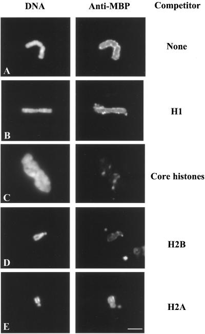
YA's binding to mitotic chromosomes can be competed by histone H2B. Binding reactions of MBP-HMK-YA with mitotic chromosomes were performed in the presence of 20 μM Drosophila histone H1 (B), equi-molar mix of Drosophila core histones (7 μM each, C), 7 μM of histone H2B (D), or 7 μM of histone H2A (E). Mitotic chromosomes were stained with DAPI (DNA) and anti-MBP antibodies (Anti-MBP). Bar: 10 μm.
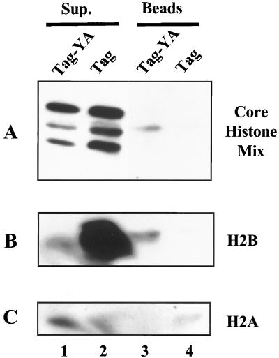
YA binds to histone H2B. MBP-HMK-YA (4 μg; Tag-YA) or MBP-HMK(32 μg; Tag) was incubated with 4 μg of a mixture of core histone (A), 4 μg histone H2B (B), or 4 μg histone H2A (C). The mixture was then incubated with amylose beads. Proteins bound to the beads were analyzed by SDS-PAGE and Western blotting with polyclonal anti-core histone antibodies (Ito et al., 1996a).
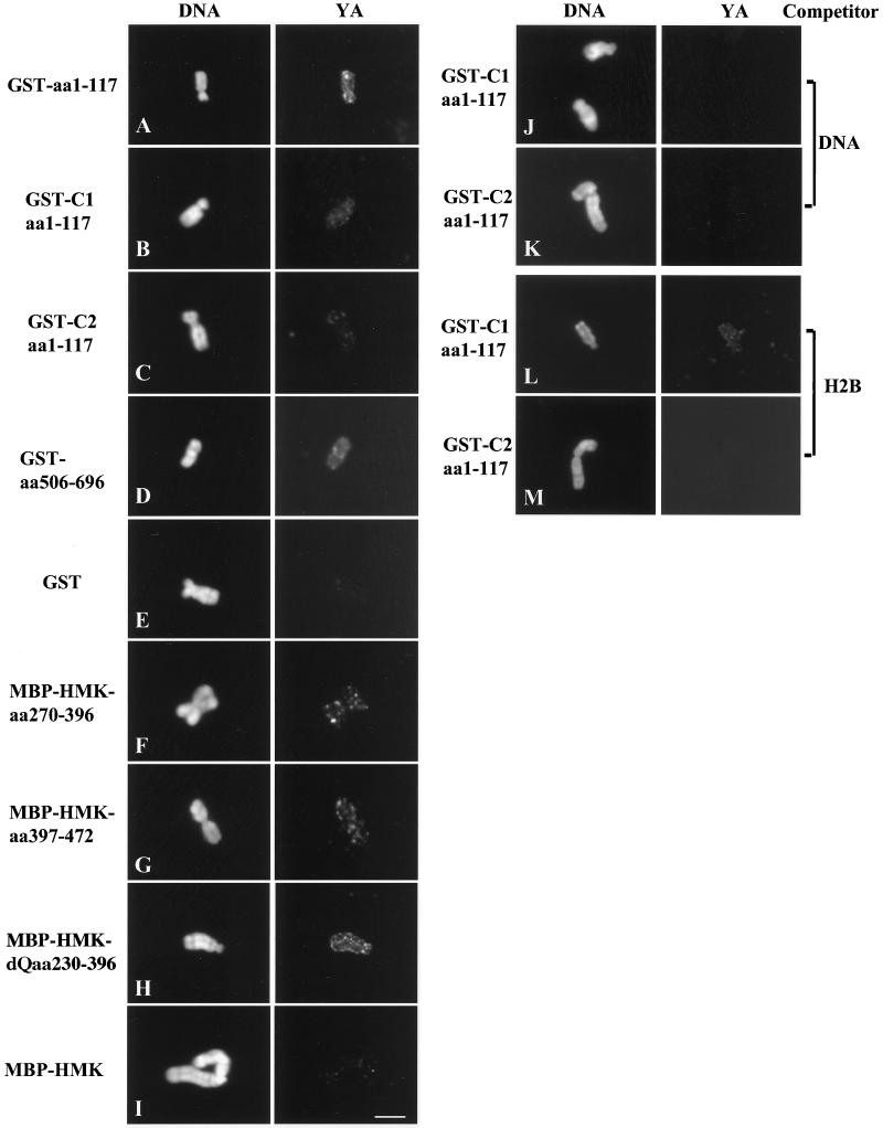
Representative YA fragments' binding to mitotic chromosomes in the presence or absence of salmon sperm DNA (DNA) or histone H2B (H2B) competitors. Purified YA fragments produced as GST or MBP-HMK fusion proteins were incubated with mitotic chromosomes. The chromosomes were stained with DAPI (DNA) and anti-GST antibodies (for GST fusion proteins, panels A–D and J–M; YA) or anti-MBP antisera (for MBP-HMK fusion proteins, panels F–H; YA). GST (panel E) or MBP-HMK (panel I) proteins were incubated with mitotic chromosomes as negative controls. Two chromosomes close to each other are shown in panel I. C1, C2: site-directed mutants in the zinc-fingers, with the cysteines in the first or the second zinc finger, respectively, mutated to alanines (Liu and Wolfner, 1998). dQ, a construct from which the glutamine-rich region was deleted. Bar: 10 μm.
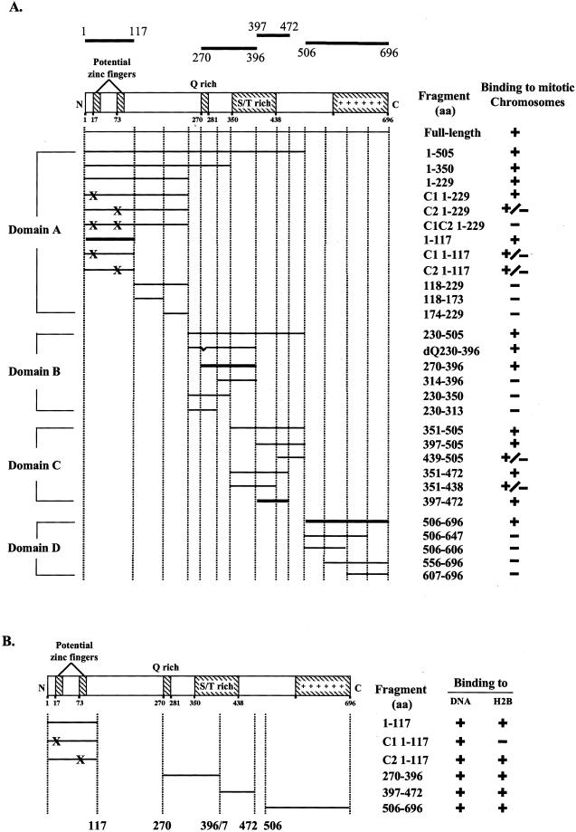
(A) Four regions in YA bind to chromosomes. Purified YA fragments as GST or MBP-HMK fusion were used for mitotic chromosome binding assays. Horizontal lines show the YA regions tested for binding. (A) Four regions, aa 1–117 (domain A), aa 270–396 (domain B), aa 397–472 (domain C), and aa 506–696 (domain D) bind to chromosomes. (B) The binding of all four regions can be competed by salmon sperm DNA and histone H2B. Q-rich: glutamine-rich region. S/T rich: Ser/Thr rich region. C1, C2, C1C2: site-directed mutants in the zinc-fingers, with the cysteines in the first, the second or both zinc fingers mutated (“X”) to alanines (Lin and Wolfner, 1991; Liu and Wolfner, 1998). dQ, a construct from which the Q-rich region was deleted. +: strong binding. +/−: very weak (close to background level of binding), −: background level of binding.
Similar articles
-
Interaction of the essential Drosophila nuclear protein YA with P0/AP3 in the cytoplasm and in vitro: implications for developmental regulation of YA's subcellular location.Dev Biol. 2002 Apr 15;244(2):429-41. doi: 10.1006/dbio.2002.0601. Dev Biol. 2002. PMID: 11944949
-
The developmentally regulated Drosophila embryonic nuclear lamina protein 'Young Arrest' (fs(1)Ya) is capable of associating with chromatin.J Cell Sci. 1997 Mar;110 ( Pt 5):643-51. doi: 10.1242/jcs.110.5.643. J Cell Sci. 1997. PMID: 9092946
-
YA is needed for proper nuclear organization to transition between meiosis and mitosis in Drosophila.BMC Dev Biol. 2009 Jul 23;9:43. doi: 10.1186/1471-213X-9-43. BMC Dev Biol. 2009. PMID: 19627584 Free PMC article.
-
[Setting the stage for mitosis: kinase regulation of prophase].Tanpakushitsu Kakusan Koso. 2004 Jun;49(8):1183-94. Tanpakushitsu Kakusan Koso. 2004. PMID: 15209214 Review. Japanese. No abstract available.
-
The right dose for every sex.Chromosoma. 2007 Apr;116(2):95-106. doi: 10.1007/s00412-006-0089-x. Epub 2006 Nov 24. Chromosoma. 2007. PMID: 17124606 Free PMC article. Review.
Cited by
-
Regulation of maternal transcript destabilization during egg activation in Drosophila.Genetics. 2003 Jul;164(3):989-1001. doi: 10.1093/genetics/164.3.989. Genetics. 2003. PMID: 12871909 Free PMC article.
References
-
- Berman CL. The Role of YA Protein in Drosophila Female Meiosis [M.S. Thesis]. Ithaca, NY: Cornell University; 2000.
-
- Blanar MA, Rutter WJ. Interaction cloning: identification of a helix-loop-helix zipper protein that interacts with c-Fos. Science. 1992;256:1014–1018. - PubMed
-
- Bulger M, Kadonaga JT. Biochemical reconstitution of chromatin with physiological nucleosome spacing. Meth Molec Genet. 1994;5:241–262.
-
- Callaini G, Riparbelli MG. Fertilization in Drosophila melanogaster: centrosome inheritance and organization of the first mitotic spindle. Dev Biol. 1996;176:199–208. - PubMed
-
- Cameron LA, Poccia DL. In vitro development of the sea urchin male pronucleus. Dev Biol. 1994;162:568–578. - PubMed
Publication types
MeSH terms
Substances
Grants and funding
LinkOut - more resources
Full Text Sources
Molecular Biology Databases
