. 2002 Feb;13(2):593-606.
doi: 10.1091/mbc.01-06-0309.

Daughter cell assembly in the protozoan parasite Toxoplasma gondii

Affiliations

Daughter cell assembly in the protozoan parasite Toxoplasma gondii

Ke Hu et al. Mol Biol Cell. 2002 Feb.

Abstract

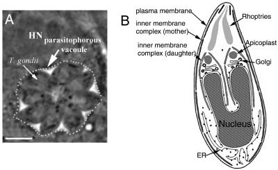
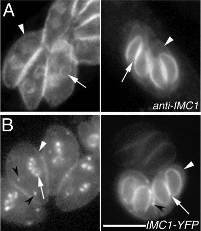
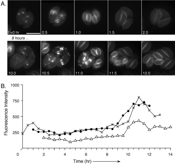
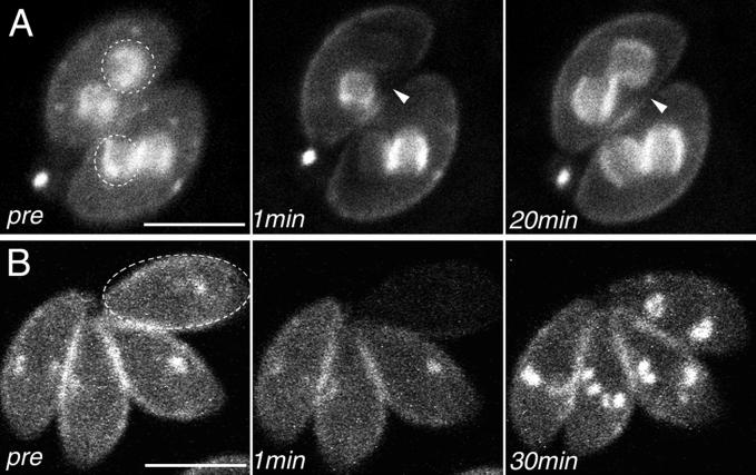
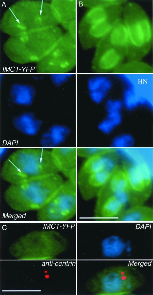
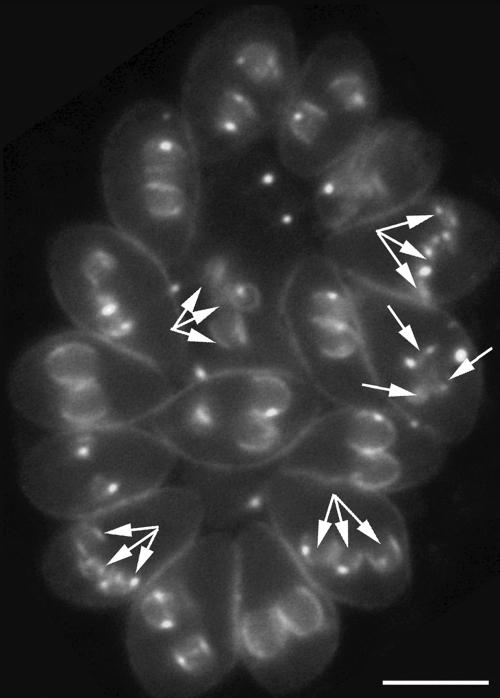
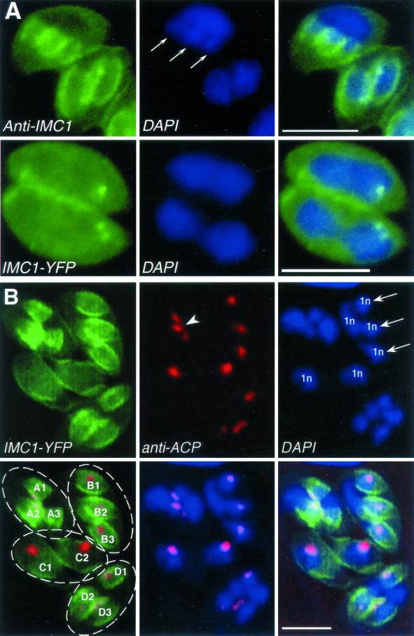
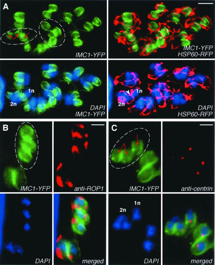
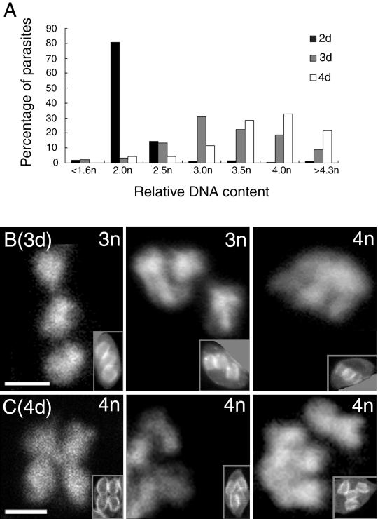
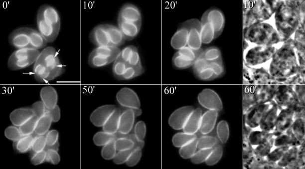
The phylum Apicomplexa includes thousands of species of obligate intracellular parasites, many of which are significant human and/or animal pathogens. Parasites in this phylum replicate by assembling daughters within the mother, using a cytoskeletal and membranous scaffolding termed the inner membrane complex. Most apicomplexan parasites, including Plasmodium sp. (which cause malaria), package many daughters within a single mother during mitosis, whereas Toxoplasma gondii typically packages only two. The comparatively simple pattern of T. gondii cell division, combined with its molecular genetic and cell biological accessibility, makes this an ideal system to study parasite cell division. A recombinant fusion between the fluorescent protein reporter YFP and the inner membrane complex protein IMC1 has been exploited to examine daughter scaffold formation in T. gondii. Time-lapse video microscopy permits the entire cell cycle of these parasites to be visualized in vivo. In addition to replication via endodyogeny (packaging two parasites at a time), T. gondii is also capable of forming multiple daughters, suggesting fundamental similarities between cell division in T. gondii and other apicomplexan parasites.


Figures

Figure 1
Figure 1
T. gondii tachyzoites establish a specialized parasitophorous vacuole inside of the host cell, within which they replicate synchronously to produce 2 parasites, 4 parasites, 8 parasites, etc. (A) Phase-contrast image showing a parasitophorous vacuole (dotted line, large arrow) containing eight T. gondii tachyzoites (small arrow). HN, host cell nucleus; bar: 5 μm. (B) Diagram of T. gondii showing the formation of daughter inner membrane complexes within the mother during cell division and the partitioning of several organelles. Note that the mother's inner membrane complex is still present at this time.
Figure 2
Figure 2
IMC1 labeling patterns of wild-type and transgenic parasites. (A) Wild-type parasites (RH) stained with IMC1 antibody during early (left) and late (right) stages of cell division. (B) Living IMC1-YFP transgenic parasites during early (left) and late (right) stages of cell division. Maternal inner membrane complexes are indicated by white arrowheads; daughter inner membrane complexes by white arrows. Note the bright concentration of fluorescence visible in IMC1-YFP transgenics (black arrowheads). Bar: 5 μm.
Figure 3
Figure 3
Time-lapse images of IMC1-YFP transgenic parasites throughout the cell division cycle. (A) Time-lapse video microscopy of IMC1-YFP transgenic parasites through two mitotic divisions over the course of 12 h. Pictures were taken at 0.5-h intervals, but only time points from 0 to 2 h and 10 to 12 h are shown, because little change was observed from 2 to 10 h; bar: 5 μm. (B) Fluorescence intensity of three parasitophorous vacuoles in a single field, corrected for background and photobleaching and plotted as a function of time. These three vacuoles were at different points of cell division at the beginning of the experiment; to facilitate comparison, curves have been shifted horizontally to align the second peak.
Figure 4
Figure 4
FRAP analysis of YFP-IMC1 incorporation and exchange. (A) One daughter (dashed circle and ellipse) in each of the two parasites in this vacuole was bleached, along with a small portion of the maternal wall (arrowheads). After 20 min, YFP fluorescence recovered uniformly in the daughters, but a defect remains visible in the maternal wall. (B) An entire mother containing two daughters at an early stage of development was bleached (dashed ellipse). After 20 min, the daughters recovered fluorescence and appear almost as bright as unbleached daughters in adjacent parasites. Bars: 5 μm.
Figure 5
Figure 5
Markers of the T. gondii cell cycle. Fixed IMC1-YFP transgenic parasites were stained with DAPI to visualize nuclear DNA (blue), and with anticentrin antibody to reveal the centrioles (red). IMC1-YFP (green) labels the inner membrane complexes. (A) Daughter scaffold formation (arrows) is initiated close to the nucleus, which remains roughly spherical at this early stage of cell division. (B) As the daughter inner membrane complexes grow in size, the dividing nucleus extends to form a horseshoe or elongated shape, depending on the orientation of the two daughters. HN, host cell nucleus. (C) Anticentrin staining reveals centriolar replication before the establishment of daughter inner membrane complexes or nuclear division. Quantitation of DAPI staining indicates that DNA replication has initiated shortly before this stage (see text). Bars: 5 μm.
Figure 6
Figure 6
Multiple cases of multiple daughter formation within mothers in a single parasitophorous vacuole. Five IMC1-YFP transgenic parasites in this parasitophorous vacuole are in the process of producing three daughters each (arrows), while 12 are producing two daughters each. The presence of 17 mothers in this vacuole suggests that one mother produced three daughters in the previous replicative cycle. Bar: 5 μm.
Figure 7
Figure 7
Nuclear and apicoplast segregation in wild-type and IMC1-YFP transgenics forming multiple daughters or nuclei. (A) Top panels: wild-type parasite stained with anti-IMC1 antibody (green) and DAPI (blue). (Note: only two of four mothers in this vacuole are shown.) The parasite in the top left corner is in the process of forming three daughters. Nuclear division produces three symmetric lobes (arrows), one for each daughter. Quantitation reveals 4n DNA content in this nucleus (see text). Lower panels: IMC1-YFP transgenic parasites (green) stained with DAPI (blue). Both parasites in this vacuole are finishing one round of nuclear division without forming daughter scaffolds (most obvious in the lower parasite), similar to what happens in schizogony. (B) IMC1-YFP transgenic parasites (green) stained with anti-ACP (a nuclear encoded apicoplast protein [Waller et al., 1998]; red) and DAPI (blue), highlighting the apicoplasts and nuclei, respectively. Four parasites are present in this vacuole (circles at lower left), and three of these (A, B, and D) are in the process of forming three daughters each. Daughter formation proceeds more rapidly when only two daughters are assembled; parasites C1 and C2 are already emerging from the mother. Note the dividing apicoplast intermediate with three symmetric branches in parasite A (arrowhead) and a nucleus in parasite B containing three lobes of 1n DNA content (arrows) and a vestigial body of 1n DNA content (see text for further description). DNA content was established by reference to the integrated DAPI fluorescence in parasites in the same or adjacent vacuole that had just finished their nuclear division. Bars: 5 μm.
Figure 8
Figure 8
Mitochondria, rhoptries, and centrioles in IMC1-YFP transgenics forming multiple daughters. Green indicates IMC1-YFP fluorescence, and blue indicates DAPI-stained DNA. Circles indicate parasites producing multiple daughters. (A) Circles indicate one parasite within this vacuole in the process of forming three daughters and another in the process of forming four daughters. Each one of the three or four daughters is associated with one nuclear lobe (or one nucleus) and one branch of the dividing mitochondrion (visualized using Hsp60-RFP; red). At this stage, one parasite contains distinct nuclei of 1n and 2n; note that daughters associating with different nuclei (antiparallel red arrows) share the same dividing mitochondrion (white arrowhead). (B) Staining with anti-ROP1 reveals that each daughter contains rhoptries. (C) One of the two parasites in this vacuole is in the process of forming three daughters (circle, red arrows). Its nucleus has divided into two nuclei (1n + 2n DNA content); the latter is in the process of further division to form a total of three daughters. Each daughter is associated with one nucleus or nuclear lobe and one centriole pair. Bars: 2 μm.
Figure 9
Figure 9
Relative DNA content among populations of parasites forming 2, 3, or 4 daughters. (A) Integrated fluorescence intensity was used to establish DNA content in individual parasite nuclei. Calibration was based on DAPI staining of neighboring parasites that had recently completed DNA replication (daughter formation well underway, but before complete emergence from the mother; see text). SE of the mean for this reference population was 0.02n. Sample sizes were as follows: 2 daughters (black), 284; 3 daughters (gray), 90; and 4 daughters (white), 35. (B) Morphological patterns exhibited by parasites in the process of forming three daughter nuclei fall into two classes consistent with either (i) progression from 1n to 2n followed by nuclear division without cytokinesis and further DNA replication in only one nucleus (center panel), or (ii) replication to 4n followed by packaging of only three daughters with 1n DNA content left behind as a residual body (right panel). (C) Morphological patterns exhibited by parasites in the process of forming four daughters are also consistent with this model, forming either two nuclei which each divide to form two daughters (center panel), or forming four daughters simultaneously from a 4n nucleus (left panel). Insets illustrate the IMC1-YFP labeling in the corresponding parasites (masked to hide other parasites in the same vacuole to facilitate interpretation). Bars: 2 μm (insets reduced by threefold).
Figure 10
Figure 10
Time-lapse video microscopy of multiple daughter formation in IMC1-YFP transgenic parasites. Replication proceeds in parallel for the three parasites that form two daughters each, and the single parasite that forms four daughters (lower right, arrows). Bar: 5 μm.

References

    1. Aikawa M. Plasmodium: the fine structure of malarial parasites. Exp Parasitol. 1971;30:284–320. - PubMed
    1. Azab M, Beverley JK. Schizogony of Toxoplasma gondii in tissue cultures. Z Parasitenkd. 1974;44:33–41. - PubMed
    1. Bannister LH, Hopkins JM, Fowler RE, Krishna S, Mitchell GH. A brief illustrated guide to the ultrastructure of Plasmodium falciparum asexual blood stages. Parasitol Today. 2000;16:427–433. - PubMed
    1. Bonhomme A, Pingret L, Pinon JM. Review: Toxoplasma gondii cellular invasion. Parassitologia. 1992;34:31–43. - PubMed
    1. Brockelman CR, Tan-Ariya P, Laovanitch R. Observation on complete schizogony of Plasmodium vivax in vitro. J Protozool. 1985;32:76–80. - PubMed

Publication types

LinkOut - more resources